Lead »
PDB 1afv-3ec8 »
2o3c »
Lead in PDB 2o3c: Crystal Structure of Zebrafish Ape
Protein crystallography data
The structure of Crystal Structure of Zebrafish Ape, PDB code: 2o3c
was solved by
M.M.Georgiadis,
R.K.Gaur,
S.Delaplane,
J.Svenson,
with X-Ray Crystallography technique. A brief refinement statistics is given in the table below:
| Resolution Low / High (Å) | 50.00 / 2.30 |
| Space group | P 1 21 1 |
| Cell size a, b, c (Å), α, β, γ (°) | 54.660, 117.780, 85.840, 90.00, 98.34, 90.00 |
| R / Rfree (%) | 19.9 / 23.6 |
Lead Binding Sites:
The binding sites of Lead atom in the Crystal Structure of Zebrafish Ape
(pdb code 2o3c). This binding sites where shown within
5.0 Angstroms radius around Lead atom.
In total 3 binding sites of Lead where determined in the Crystal Structure of Zebrafish Ape, PDB code: 2o3c:
Jump to Lead binding site number: 1; 2; 3;
In total 3 binding sites of Lead where determined in the Crystal Structure of Zebrafish Ape, PDB code: 2o3c:
Jump to Lead binding site number: 1; 2; 3;
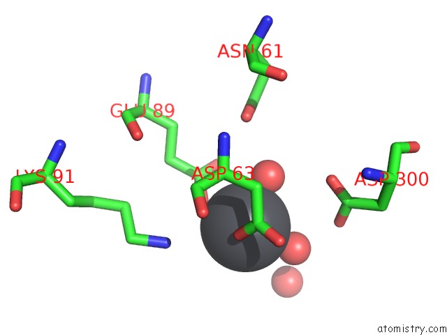
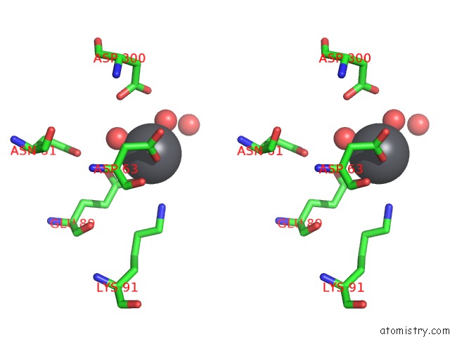
Lead binding site 1 out of 3 in 2o3c
Go back to
Lead binding site 1 out
of 3 in the Crystal Structure of Zebrafish Ape
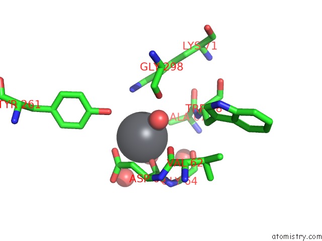
Mono view
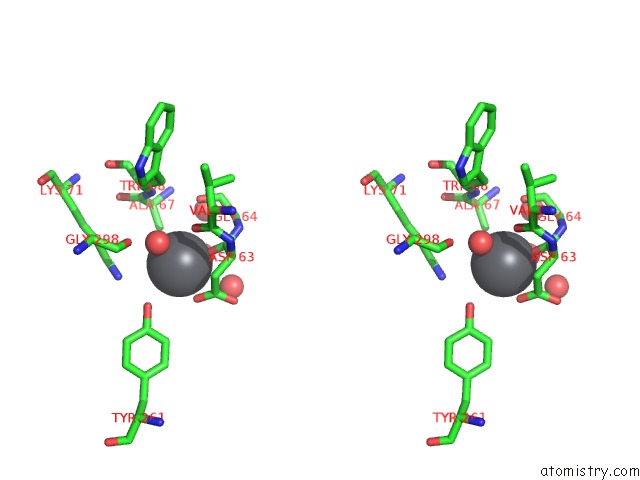
Stereo pair view
Mono view
Stereo pair view
A full contact list of Lead with other atoms in the Pb binding
site number 1 of Crystal Structure of Zebrafish Ape within 5.0Å range:
|
Lead binding site 2 out of 3 in 2o3c
Go back to
Lead binding site 2 out
of 3 in the Crystal Structure of Zebrafish Ape
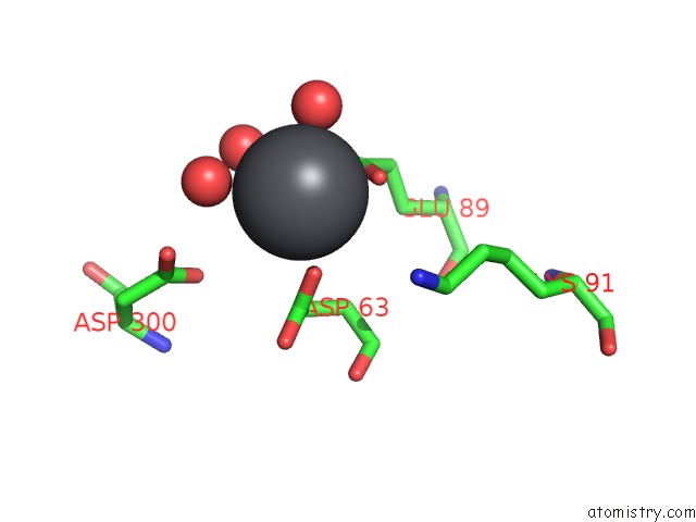
Mono view
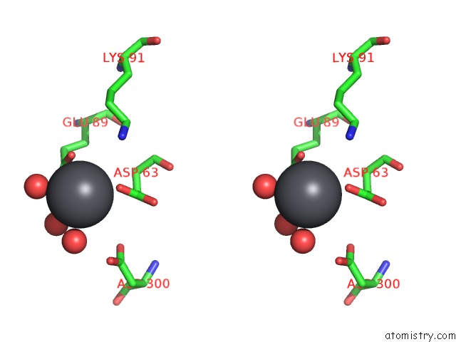
Stereo pair view
Mono view
Stereo pair view
A full contact list of Lead with other atoms in the Pb binding
site number 2 of Crystal Structure of Zebrafish Ape within 5.0Å range:
|
Lead binding site 3 out of 3 in 2o3c
Go back to
Lead binding site 3 out
of 3 in the Crystal Structure of Zebrafish Ape
Mono view
Stereo pair view
Mono view
Stereo pair view
A full contact list of Lead with other atoms in the Pb binding
site number 3 of Crystal Structure of Zebrafish Ape within 5.0Å range:
|
Reference:
M.M.Georgiadis,
M.Luo,
R.K.Gaur,
S.Delaplane,
X.Li,
M.R.Kelley.
Evolution of the Redox Function in Mammalian Apurinic/Apyrimidinic Endonuclease Mutat.Res. V. 643 54 2008.
ISSN: ISSN 0027-5107
PubMed: 18579163
DOI: 10.1016/J.MRFMMM.2008.04.008
Page generated: Thu Oct 10 10:04:54 2024
ISSN: ISSN 0027-5107
PubMed: 18579163
DOI: 10.1016/J.MRFMMM.2008.04.008
Last articles
K in 4KM7K in 4KMY
K in 4KMX
K in 4KI2
K in 4KFM
K in 4KKY
K in 4KGD
K in 4KCW
K in 4KCT
K in 4KCV