Lead »
PDB 1afv-3ec8 »
1sn8 »
Lead in PDB 1sn8: Crystal Structure of the S1 Domain of Rnase E From E. Coli (Pb Derivative)
Protein crystallography data
The structure of Crystal Structure of the S1 Domain of Rnase E From E. Coli (Pb Derivative), PDB code: 1sn8
was solved by
M.Schubert,
R.E.Edge,
P.Lario,
M.A.Cook,
N.C.J.Strynadka,
G.A.Mackie,
L.P.Mcintosh,
with X-Ray Crystallography technique. A brief refinement statistics is given in the table below:
| Resolution Low / High (Å) | 25.00 / 2.00 |
| Space group | P 41 21 2 |
| Cell size a, b, c (Å), α, β, γ (°) | 70.580, 70.580, 87.902, 90.00, 90.00, 90.00 |
| R / Rfree (%) | 18.2 / 23.2 |
Lead Binding Sites:
The binding sites of Lead atom in the Crystal Structure of the S1 Domain of Rnase E From E. Coli (Pb Derivative)
(pdb code 1sn8). This binding sites where shown within
5.0 Angstroms radius around Lead atom.
In total 2 binding sites of Lead where determined in the Crystal Structure of the S1 Domain of Rnase E From E. Coli (Pb Derivative), PDB code: 1sn8:
Jump to Lead binding site number: 1; 2;
In total 2 binding sites of Lead where determined in the Crystal Structure of the S1 Domain of Rnase E From E. Coli (Pb Derivative), PDB code: 1sn8:
Jump to Lead binding site number: 1; 2;
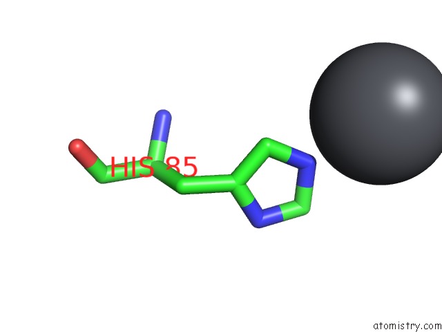
Lead binding site 1 out of 2 in 1sn8
Go back to
Lead binding site 1 out
of 2 in the Crystal Structure of the S1 Domain of Rnase E From E. Coli (Pb Derivative)
Mono view
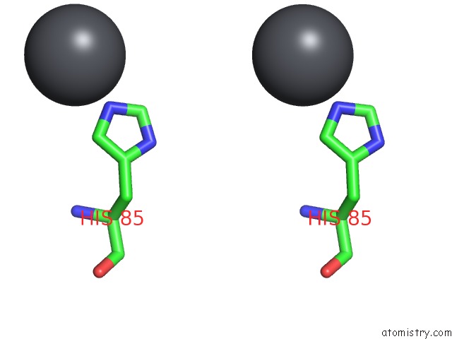
Stereo pair view
Mono view
Stereo pair view
A full contact list of Lead with other atoms in the Pb binding
site number 1 of Crystal Structure of the S1 Domain of Rnase E From E. Coli (Pb Derivative) within 5.0Å range:
|
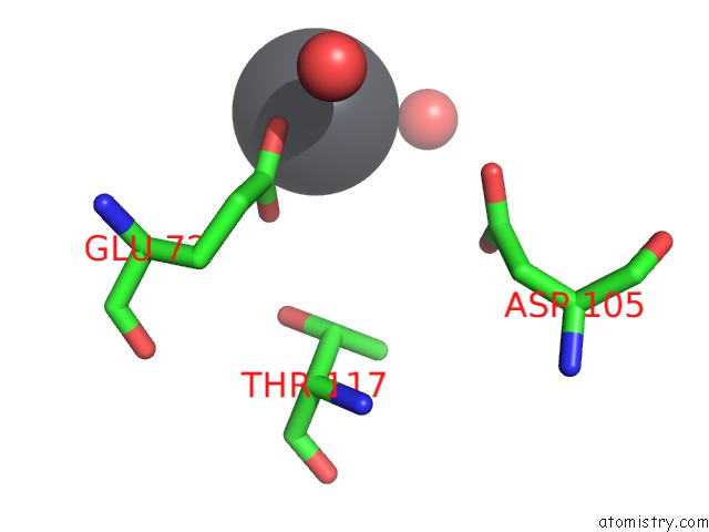
Lead binding site 2 out of 2 in 1sn8
Go back to
Lead binding site 2 out
of 2 in the Crystal Structure of the S1 Domain of Rnase E From E. Coli (Pb Derivative)
Mono view
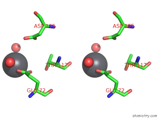
Stereo pair view
Mono view
Stereo pair view
A full contact list of Lead with other atoms in the Pb binding
site number 2 of Crystal Structure of the S1 Domain of Rnase E From E. Coli (Pb Derivative) within 5.0Å range:
|
Reference:
M.Schubert,
R.E.Edge,
P.Lario,
M.A.Cook,
N.C.Strynadka,
G.A.Mackie,
L.P.Mcintosh.
Structural Characterization of the Rnase E S1 Domain and Identification of Its Oligonucleotide-Binding and Dimerization Interfaces. J.Mol.Biol. V. 341 37 2004.
ISSN: ISSN 0022-2836
PubMed: 15312761
DOI: 10.1016/J.JMB.2004.05.061
Page generated: Thu Oct 10 10:02:13 2024
ISSN: ISSN 0022-2836
PubMed: 15312761
DOI: 10.1016/J.JMB.2004.05.061
Last articles
K in 8K1SK in 8JXP
K in 8JXO
K in 8K1Q
K in 8K1J
K in 8K1E
K in 8JZG
K in 8K0U
K in 8K0T
K in 8JGW