Lead »
PDB 1afv-3ec8 »
1qr7 »
Lead in PDB 1qr7: Crystal Structure of Phenylalanine-Regulated 3-Deoxy-D- Arabino-Heptulosonate-7-Phosphate Synthase From Escherichia Coli Complexed with PB2+ and Pep
Enzymatic activity of Crystal Structure of Phenylalanine-Regulated 3-Deoxy-D- Arabino-Heptulosonate-7-Phosphate Synthase From Escherichia Coli Complexed with PB2+ and Pep
All present enzymatic activity of Crystal Structure of Phenylalanine-Regulated 3-Deoxy-D- Arabino-Heptulosonate-7-Phosphate Synthase From Escherichia Coli Complexed with PB2+ and Pep:
4.1.2.15;
4.1.2.15;
Protein crystallography data
The structure of Crystal Structure of Phenylalanine-Regulated 3-Deoxy-D- Arabino-Heptulosonate-7-Phosphate Synthase From Escherichia Coli Complexed with PB2+ and Pep, PDB code: 1qr7
was solved by
I.A.Shumilin,
R.H.Kretsinger,
R.H.Bauerle,
with X-Ray Crystallography technique. A brief refinement statistics is given in the table below:
| Resolution Low / High (Å) | 18.00 / 2.60 |
| Space group | C 1 2 1 |
| Cell size a, b, c (Å), α, β, γ (°) | 211.731, 51.326, 148.099, 90.00, 116.43, 90.00 |
| R / Rfree (%) | 19.4 / 25.6 |
Lead Binding Sites:
The binding sites of Lead atom in the Crystal Structure of Phenylalanine-Regulated 3-Deoxy-D- Arabino-Heptulosonate-7-Phosphate Synthase From Escherichia Coli Complexed with PB2+ and Pep
(pdb code 1qr7). This binding sites where shown within
5.0 Angstroms radius around Lead atom.
In total 4 binding sites of Lead where determined in the Crystal Structure of Phenylalanine-Regulated 3-Deoxy-D- Arabino-Heptulosonate-7-Phosphate Synthase From Escherichia Coli Complexed with PB2+ and Pep, PDB code: 1qr7:
Jump to Lead binding site number: 1; 2; 3; 4;
In total 4 binding sites of Lead where determined in the Crystal Structure of Phenylalanine-Regulated 3-Deoxy-D- Arabino-Heptulosonate-7-Phosphate Synthase From Escherichia Coli Complexed with PB2+ and Pep, PDB code: 1qr7:
Jump to Lead binding site number: 1; 2; 3; 4;
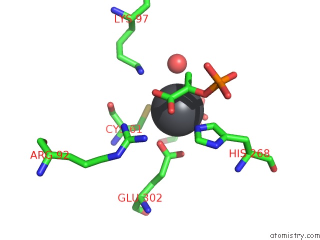
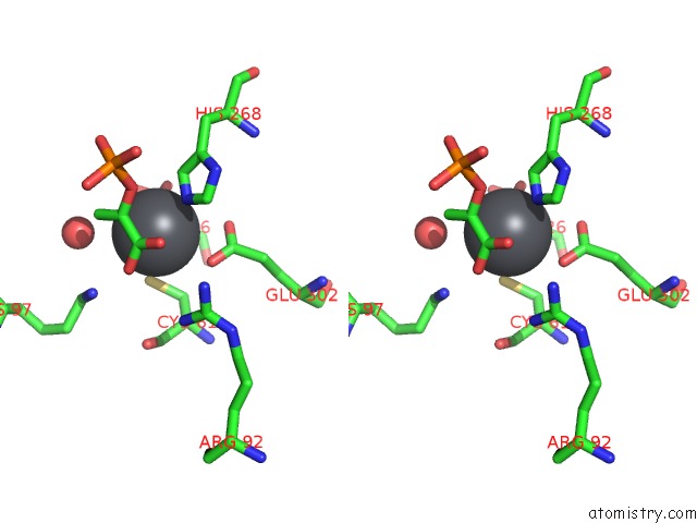
Lead binding site 1 out of 4 in 1qr7
Go back to
Lead binding site 1 out
of 4 in the Crystal Structure of Phenylalanine-Regulated 3-Deoxy-D- Arabino-Heptulosonate-7-Phosphate Synthase From Escherichia Coli Complexed with PB2+ and Pep
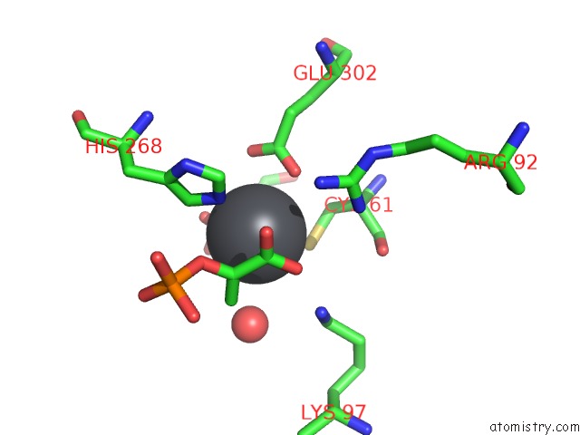
Mono view
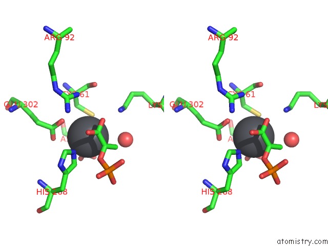
Stereo pair view
Mono view
Stereo pair view
A full contact list of Lead with other atoms in the Pb binding
site number 1 of Crystal Structure of Phenylalanine-Regulated 3-Deoxy-D- Arabino-Heptulosonate-7-Phosphate Synthase From Escherichia Coli Complexed with PB2+ and Pep within 5.0Å range:
|
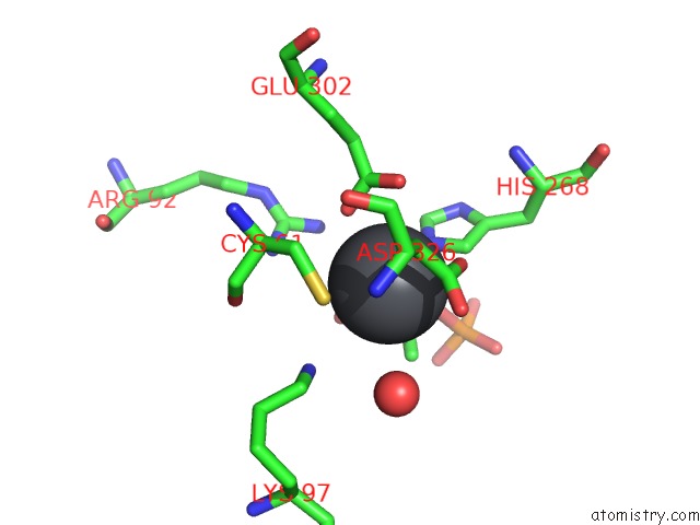
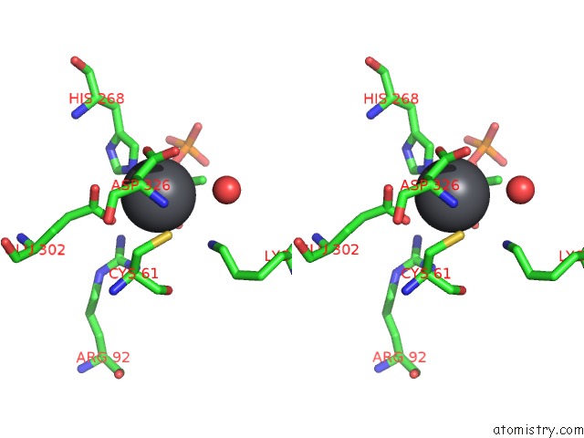
Lead binding site 2 out of 4 in 1qr7
Go back to
Lead binding site 2 out
of 4 in the Crystal Structure of Phenylalanine-Regulated 3-Deoxy-D- Arabino-Heptulosonate-7-Phosphate Synthase From Escherichia Coli Complexed with PB2+ and Pep
Mono view
Stereo pair view
Mono view
Stereo pair view
A full contact list of Lead with other atoms in the Pb binding
site number 2 of Crystal Structure of Phenylalanine-Regulated 3-Deoxy-D- Arabino-Heptulosonate-7-Phosphate Synthase From Escherichia Coli Complexed with PB2+ and Pep within 5.0Å range:
|
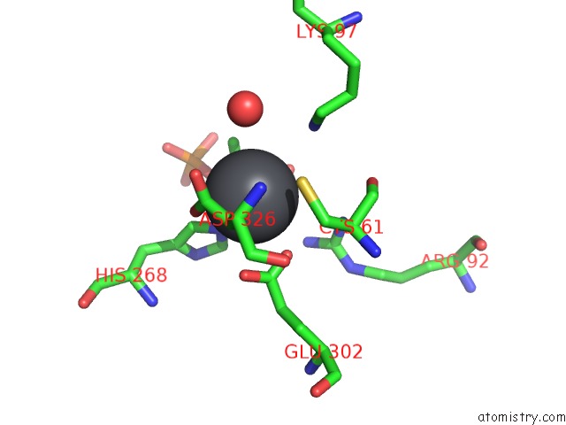
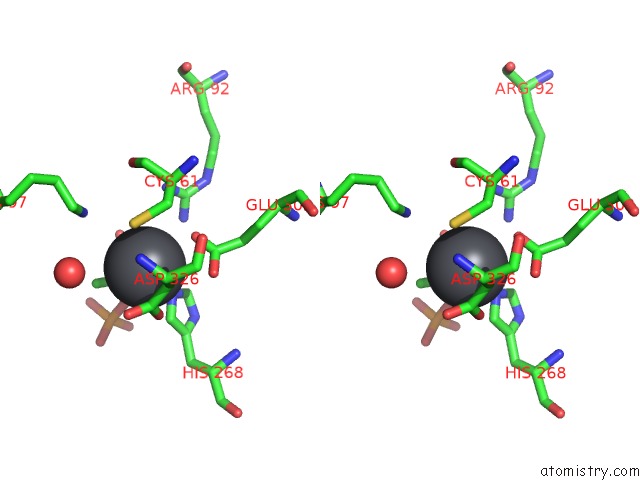
Lead binding site 3 out of 4 in 1qr7
Go back to
Lead binding site 3 out
of 4 in the Crystal Structure of Phenylalanine-Regulated 3-Deoxy-D- Arabino-Heptulosonate-7-Phosphate Synthase From Escherichia Coli Complexed with PB2+ and Pep
Mono view
Stereo pair view
Mono view
Stereo pair view
A full contact list of Lead with other atoms in the Pb binding
site number 3 of Crystal Structure of Phenylalanine-Regulated 3-Deoxy-D- Arabino-Heptulosonate-7-Phosphate Synthase From Escherichia Coli Complexed with PB2+ and Pep within 5.0Å range:
|
Lead binding site 4 out of 4 in 1qr7
Go back to
Lead binding site 4 out
of 4 in the Crystal Structure of Phenylalanine-Regulated 3-Deoxy-D- Arabino-Heptulosonate-7-Phosphate Synthase From Escherichia Coli Complexed with PB2+ and Pep
Mono view
Stereo pair view
Mono view
Stereo pair view
A full contact list of Lead with other atoms in the Pb binding
site number 4 of Crystal Structure of Phenylalanine-Regulated 3-Deoxy-D- Arabino-Heptulosonate-7-Phosphate Synthase From Escherichia Coli Complexed with PB2+ and Pep within 5.0Å range:
|
Reference:
I.A.Shumilin,
R.H.Kretsinger,
R.H.Bauerle.
Crystal Structure of Phenylalanine-Regulated 3-Deoxy-D-Arabino-Heptulosonate-7-Phosphate Synthase From Escherichia Coli. Structure Fold.Des. V. 7 865 1999.
ISSN: ISSN 0969-2126
PubMed: 10425687
DOI: 10.1016/S0969-2126(99)80109-9
Page generated: Thu Oct 10 10:01:59 2024
ISSN: ISSN 0969-2126
PubMed: 10425687
DOI: 10.1016/S0969-2126(99)80109-9
Last articles
K in 4U69K in 4TWK
K in 4TS2
K in 4TOH
K in 4TOE
K in 4TOG
K in 4TOF
K in 4TOD
K in 4TOA
K in 4TO9