Lead »
PDB 1afv-3ec8 »
1nbs »
Lead in PDB 1nbs: Crystal Structure of the Specificity Domain of Ribonuclease P Rna
Protein crystallography data
The structure of Crystal Structure of the Specificity Domain of Ribonuclease P Rna, PDB code: 1nbs
was solved by
A.S.Krasilnikov,
X.Yang,
T.Pan,
A.Mondragon,
with X-Ray Crystallography technique. A brief refinement statistics is given in the table below:
| Resolution Low / High (Å) | 33.00 / 3.15 |
| Space group | C 2 2 21 |
| Cell size a, b, c (Å), α, β, γ (°) | 126.500, 145.300, 144.600, 90.00, 90.00, 90.00 |
| R / Rfree (%) | 28 / 30.7 |
Other elements in 1nbs:
The structure of Crystal Structure of the Specificity Domain of Ribonuclease P Rna also contains other interesting chemical elements:
| Magnesium | (Mg) | 12 atoms |
Lead Binding Sites:
Pages:
>>> Page 1 <<< Page 2, Binding sites: 11 - 20; Page 3, Binding sites: 21 - 23;Binding sites:
The binding sites of Lead atom in the Crystal Structure of the Specificity Domain of Ribonuclease P Rna (pdb code 1nbs). This binding sites where shown within 5.0 Angstroms radius around Lead atom.In total 23 binding sites of Lead where determined in the Crystal Structure of the Specificity Domain of Ribonuclease P Rna, PDB code: 1nbs:
Jump to Lead binding site number: 1; 2; 3; 4; 5; 6; 7; 8; 9; 10;
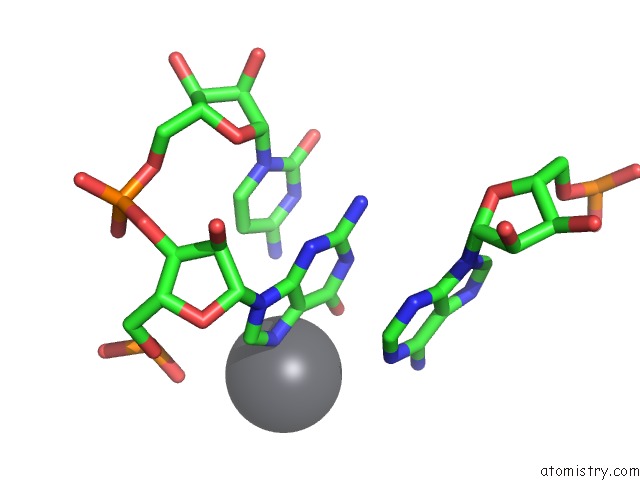
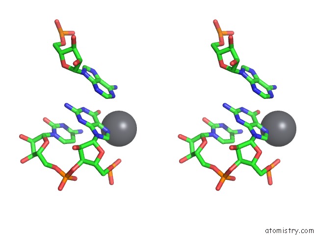
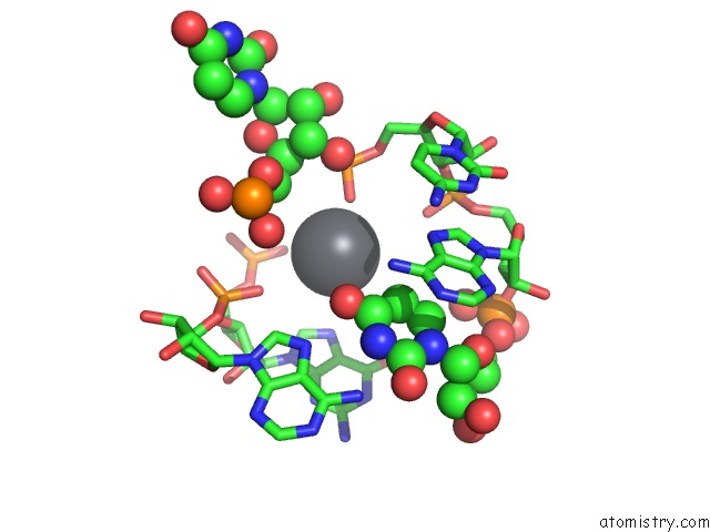
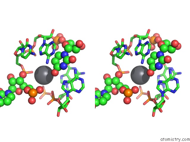
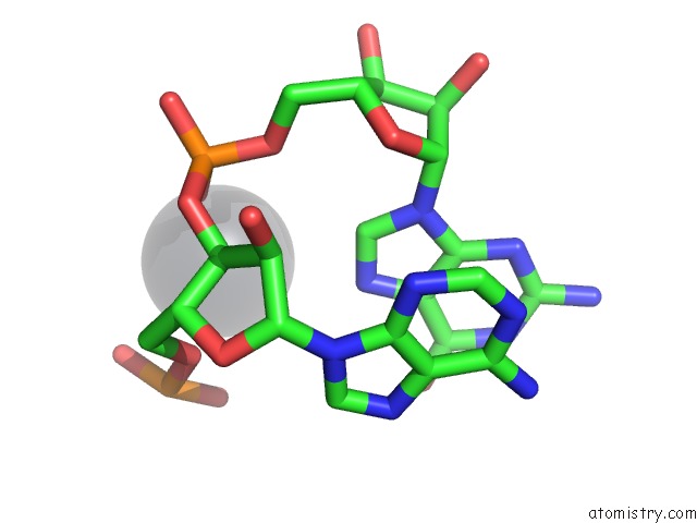
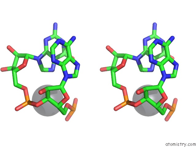
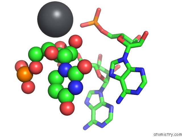
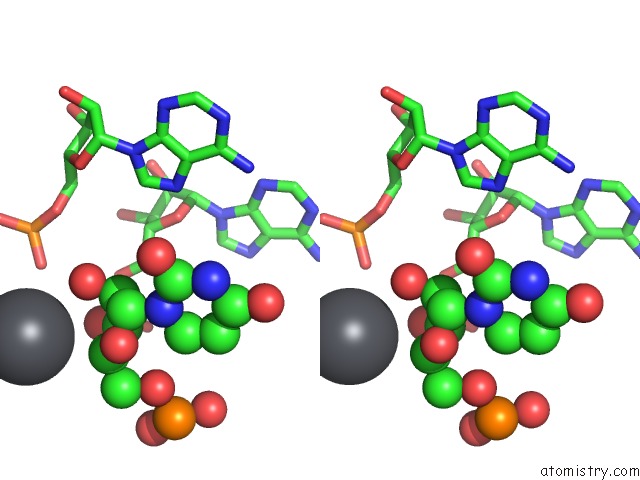
Lead binding site 1 out of 23 in 1nbs
Go back to
Lead binding site 1 out
of 23 in the Crystal Structure of the Specificity Domain of Ribonuclease P Rna
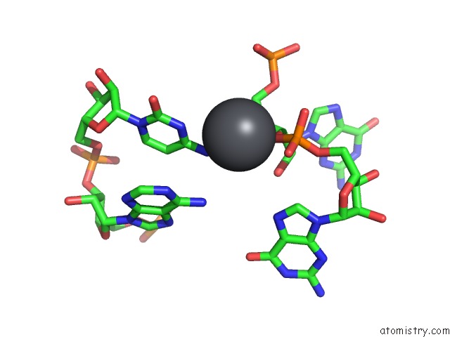
Mono view
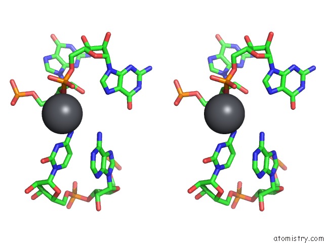
Stereo pair view
Mono view
Stereo pair view
A full contact list of Lead with other atoms in the Pb binding
site number 1 of Crystal Structure of the Specificity Domain of Ribonuclease P Rna within 5.0Å range:
|
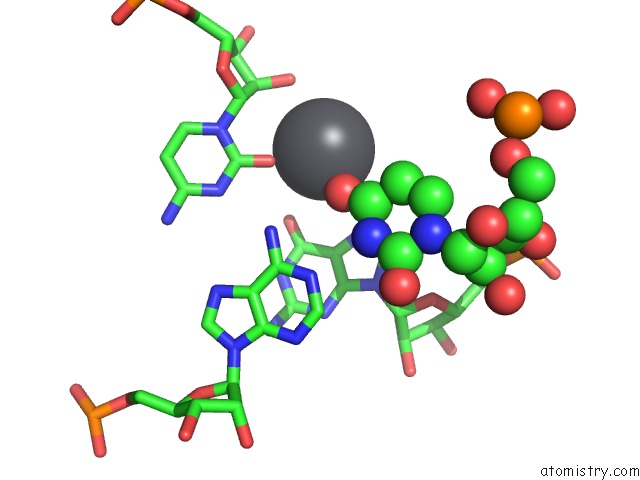
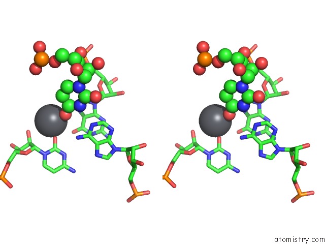
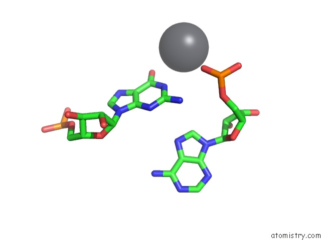
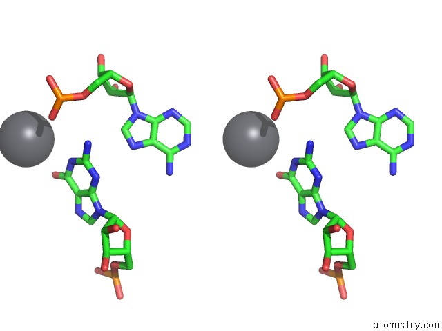
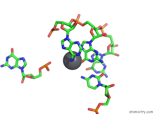
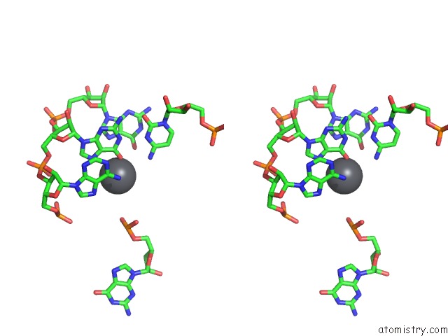
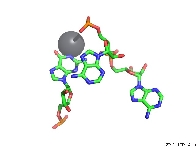
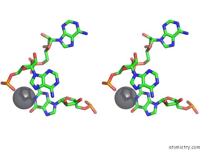
Lead binding site 2 out of 23 in 1nbs
Go back to
Lead binding site 2 out
of 23 in the Crystal Structure of the Specificity Domain of Ribonuclease P Rna
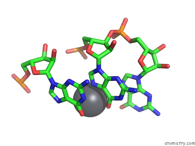
Mono view
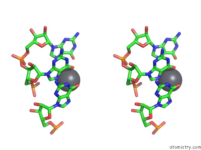
Stereo pair view
Mono view
Stereo pair view
A full contact list of Lead with other atoms in the Pb binding
site number 2 of Crystal Structure of the Specificity Domain of Ribonuclease P Rna within 5.0Å range:
|
Lead binding site 3 out of 23 in 1nbs
Go back to
Lead binding site 3 out
of 23 in the Crystal Structure of the Specificity Domain of Ribonuclease P Rna
Mono view
Stereo pair view
Mono view
Stereo pair view
A full contact list of Lead with other atoms in the Pb binding
site number 3 of Crystal Structure of the Specificity Domain of Ribonuclease P Rna within 5.0Å range:
|
Lead binding site 4 out of 23 in 1nbs
Go back to
Lead binding site 4 out
of 23 in the Crystal Structure of the Specificity Domain of Ribonuclease P Rna
Mono view
Stereo pair view
Mono view
Stereo pair view
A full contact list of Lead with other atoms in the Pb binding
site number 4 of Crystal Structure of the Specificity Domain of Ribonuclease P Rna within 5.0Å range:
|
Lead binding site 5 out of 23 in 1nbs
Go back to
Lead binding site 5 out
of 23 in the Crystal Structure of the Specificity Domain of Ribonuclease P Rna
Mono view
Stereo pair view
Mono view
Stereo pair view
A full contact list of Lead with other atoms in the Pb binding
site number 5 of Crystal Structure of the Specificity Domain of Ribonuclease P Rna within 5.0Å range:
|
Lead binding site 6 out of 23 in 1nbs
Go back to
Lead binding site 6 out
of 23 in the Crystal Structure of the Specificity Domain of Ribonuclease P Rna
Mono view
Stereo pair view
Mono view
Stereo pair view
A full contact list of Lead with other atoms in the Pb binding
site number 6 of Crystal Structure of the Specificity Domain of Ribonuclease P Rna within 5.0Å range:
|
Lead binding site 7 out of 23 in 1nbs
Go back to
Lead binding site 7 out
of 23 in the Crystal Structure of the Specificity Domain of Ribonuclease P Rna
Mono view
Stereo pair view
Mono view
Stereo pair view
A full contact list of Lead with other atoms in the Pb binding
site number 7 of Crystal Structure of the Specificity Domain of Ribonuclease P Rna within 5.0Å range:
|
Lead binding site 8 out of 23 in 1nbs
Go back to
Lead binding site 8 out
of 23 in the Crystal Structure of the Specificity Domain of Ribonuclease P Rna
Mono view
Stereo pair view
Mono view
Stereo pair view
A full contact list of Lead with other atoms in the Pb binding
site number 8 of Crystal Structure of the Specificity Domain of Ribonuclease P Rna within 5.0Å range:
|
Lead binding site 9 out of 23 in 1nbs
Go back to
Lead binding site 9 out
of 23 in the Crystal Structure of the Specificity Domain of Ribonuclease P Rna
Mono view
Stereo pair view
Mono view
Stereo pair view
A full contact list of Lead with other atoms in the Pb binding
site number 9 of Crystal Structure of the Specificity Domain of Ribonuclease P Rna within 5.0Å range:
|
Lead binding site 10 out of 23 in 1nbs
Go back to
Lead binding site 10 out
of 23 in the Crystal Structure of the Specificity Domain of Ribonuclease P Rna
Mono view
Stereo pair view
Mono view
Stereo pair view
A full contact list of Lead with other atoms in the Pb binding
site number 10 of Crystal Structure of the Specificity Domain of Ribonuclease P Rna within 5.0Å range:
|
Reference:
A.S.Krasilnikov,
X.Yang,
T.Pan,
A.Mondragon.
Crystal Structure of the Specificity Domain of Ribonuclease P Nature V. 421 760 2003.
ISSN: ISSN 0028-0836
PubMed: 12610630
DOI: 10.1038/NATURE01386
Page generated: Thu Oct 10 10:00:40 2024
ISSN: ISSN 0028-0836
PubMed: 12610630
DOI: 10.1038/NATURE01386
Last articles
Na in 1S3XNa in 1S36
Na in 1S0A
Na in 1S1K
Na in 1S09
Na in 1RZT
Na in 1S08
Na in 1S07
Na in 1S06
Na in 1RYS