Lead »
PDB 1afv-3ec8 »
1hqj »
Lead in PDB 1hqj: Crystal Structure of A De Novo Designed Trimeric Coiled-Coil Peptide
Protein crystallography data
The structure of Crystal Structure of A De Novo Designed Trimeric Coiled-Coil Peptide, PDB code: 1hqj
was solved by
P.Burkhard,
M.Meier,
A.Lustig,
with X-Ray Crystallography technique. A brief refinement statistics is given in the table below:
| Resolution Low / High (Å) | 10.00 / 1.20 |
| Space group | P 21 21 21 |
| Cell size a, b, c (Å), α, β, γ (°) | 44.334, 44.874, 81.043, 90.00, 90.00, 90.00 |
| R / Rfree (%) | n/a / 22.3 |
Lead Binding Sites:
The binding sites of Lead atom in the Crystal Structure of A De Novo Designed Trimeric Coiled-Coil Peptide
(pdb code 1hqj). This binding sites where shown within
5.0 Angstroms radius around Lead atom.
In total 9 binding sites of Lead where determined in the Crystal Structure of A De Novo Designed Trimeric Coiled-Coil Peptide, PDB code: 1hqj:
Jump to Lead binding site number: 1; 2; 3; 4; 5; 6; 7; 8; 9;
In total 9 binding sites of Lead where determined in the Crystal Structure of A De Novo Designed Trimeric Coiled-Coil Peptide, PDB code: 1hqj:
Jump to Lead binding site number: 1; 2; 3; 4; 5; 6; 7; 8; 9;
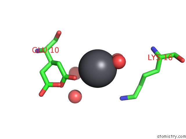
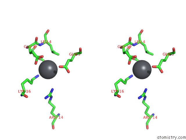
Lead binding site 1 out of 9 in 1hqj
Go back to
Lead binding site 1 out
of 9 in the Crystal Structure of A De Novo Designed Trimeric Coiled-Coil Peptide
Mono view

Stereo pair view
Mono view
Stereo pair view
A full contact list of Lead with other atoms in the Pb binding
site number 1 of Crystal Structure of A De Novo Designed Trimeric Coiled-Coil Peptide within 5.0Å range:
|
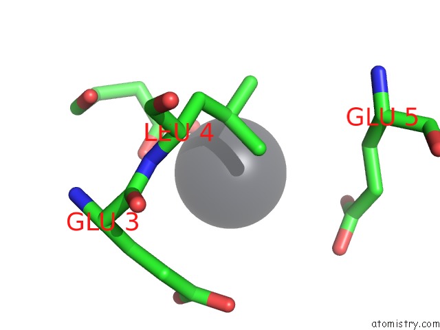
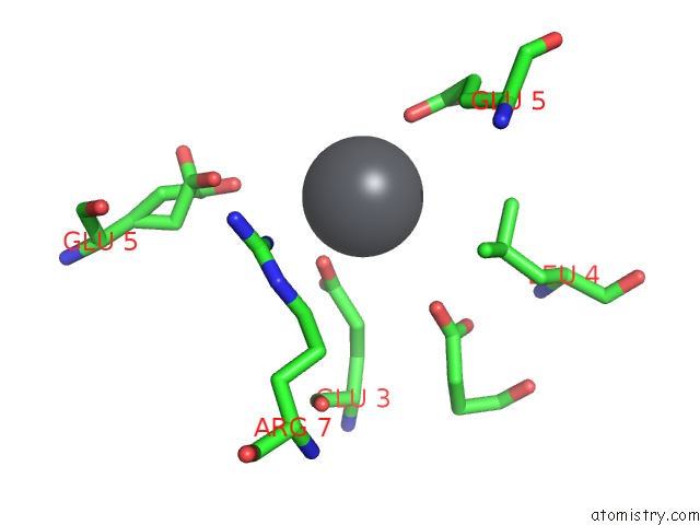
Lead binding site 2 out of 9 in 1hqj
Go back to
Lead binding site 2 out
of 9 in the Crystal Structure of A De Novo Designed Trimeric Coiled-Coil Peptide
Mono view

Stereo pair view
Mono view
Stereo pair view
A full contact list of Lead with other atoms in the Pb binding
site number 2 of Crystal Structure of A De Novo Designed Trimeric Coiled-Coil Peptide within 5.0Å range:
|
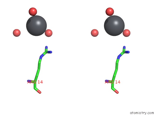
Lead binding site 3 out of 9 in 1hqj
Go back to
Lead binding site 3 out
of 9 in the Crystal Structure of A De Novo Designed Trimeric Coiled-Coil Peptide

Mono view

Stereo pair view
Mono view
Stereo pair view
A full contact list of Lead with other atoms in the Pb binding
site number 3 of Crystal Structure of A De Novo Designed Trimeric Coiled-Coil Peptide within 5.0Å range:
|
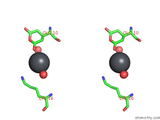
Lead binding site 4 out of 9 in 1hqj
Go back to
Lead binding site 4 out
of 9 in the Crystal Structure of A De Novo Designed Trimeric Coiled-Coil Peptide

Mono view

Stereo pair view
Mono view
Stereo pair view
A full contact list of Lead with other atoms in the Pb binding
site number 4 of Crystal Structure of A De Novo Designed Trimeric Coiled-Coil Peptide within 5.0Å range:
|
Lead binding site 5 out of 9 in 1hqj
Go back to
Lead binding site 5 out
of 9 in the Crystal Structure of A De Novo Designed Trimeric Coiled-Coil Peptide

Mono view
Stereo pair view
Mono view
Stereo pair view
A full contact list of Lead with other atoms in the Pb binding
site number 5 of Crystal Structure of A De Novo Designed Trimeric Coiled-Coil Peptide within 5.0Å range:
|
Lead binding site 6 out of 9 in 1hqj
Go back to
Lead binding site 6 out
of 9 in the Crystal Structure of A De Novo Designed Trimeric Coiled-Coil Peptide

Mono view

Stereo pair view
Mono view
Stereo pair view
A full contact list of Lead with other atoms in the Pb binding
site number 6 of Crystal Structure of A De Novo Designed Trimeric Coiled-Coil Peptide within 5.0Å range:
|
Lead binding site 7 out of 9 in 1hqj
Go back to
Lead binding site 7 out
of 9 in the Crystal Structure of A De Novo Designed Trimeric Coiled-Coil Peptide

Mono view
Stereo pair view
Mono view
Stereo pair view
A full contact list of Lead with other atoms in the Pb binding
site number 7 of Crystal Structure of A De Novo Designed Trimeric Coiled-Coil Peptide within 5.0Å range:
|
Lead binding site 8 out of 9 in 1hqj
Go back to
Lead binding site 8 out
of 9 in the Crystal Structure of A De Novo Designed Trimeric Coiled-Coil Peptide

Mono view
Stereo pair view
Mono view
Stereo pair view
A full contact list of Lead with other atoms in the Pb binding
site number 8 of Crystal Structure of A De Novo Designed Trimeric Coiled-Coil Peptide within 5.0Å range:
|
Lead binding site 9 out of 9 in 1hqj
Go back to
Lead binding site 9 out
of 9 in the Crystal Structure of A De Novo Designed Trimeric Coiled-Coil Peptide
Mono view

Stereo pair view
Mono view
Stereo pair view
A full contact list of Lead with other atoms in the Pb binding
site number 9 of Crystal Structure of A De Novo Designed Trimeric Coiled-Coil Peptide within 5.0Å range:
|
Reference:
P.Burkhard,
M.Meier,
A.Lustig.
Design of A Minimal Protein Oligomerization Domain By A Structural Approach. Protein Sci. V. 9 2294 2000.
ISSN: ISSN 0961-8368
PubMed: 11206050
Page generated: Thu Oct 10 09:59:35 2024
ISSN: ISSN 0961-8368
PubMed: 11206050
Last articles
K in 5LI1K in 5LF3
K in 5LF1
K in 5LF0
K in 5LEZ
K in 5LEY
K in 5LEX
K in 5LE5
K in 5KX9
K in 5KUK