Lead »
PDB 3fhh-8gbm »
7d33 »
Lead in PDB 7d33: The PB2+ Complexed Structure of Tba G8C Mutant
Protein crystallography data
The structure of The PB2+ Complexed Structure of Tba G8C Mutant, PDB code: 7d33
was solved by
H.H.Liu,
Y.Q.Gao,
J.Sheng,
J.H.Gan,
with X-Ray Crystallography technique. A brief refinement statistics is given in the table below:
| Resolution Low / High (Å) | 25.34 / 2.12 |
| Space group | C 1 2 1 |
| Cell size a, b, c (Å), α, β, γ (°) | 78.765, 51.941, 57.063, 90, 113.59, 90 |
| R / Rfree (%) | 21.2 / 24 |
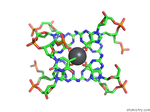
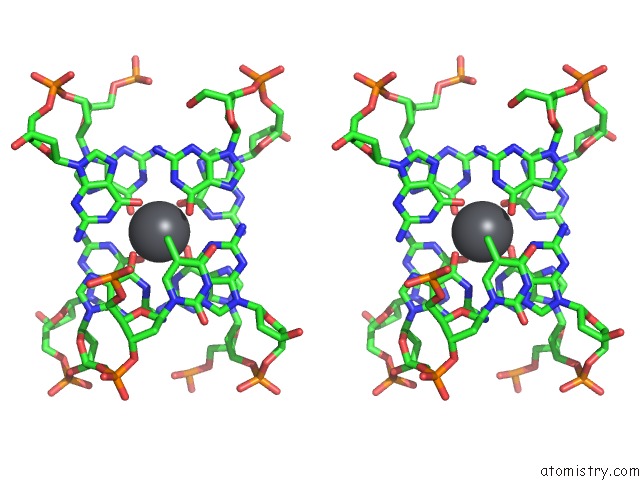
Lead Binding Sites:
The binding sites of Lead atom in the The PB2+ Complexed Structure of Tba G8C Mutant
(pdb code 7d33). This binding sites where shown within
5.0 Angstroms radius around Lead atom.
In total 5 binding sites of Lead where determined in the The PB2+ Complexed Structure of Tba G8C Mutant, PDB code: 7d33:
Jump to Lead binding site number: 1; 2; 3; 4; 5;
In total 5 binding sites of Lead where determined in the The PB2+ Complexed Structure of Tba G8C Mutant, PDB code: 7d33:
Jump to Lead binding site number: 1; 2; 3; 4; 5;
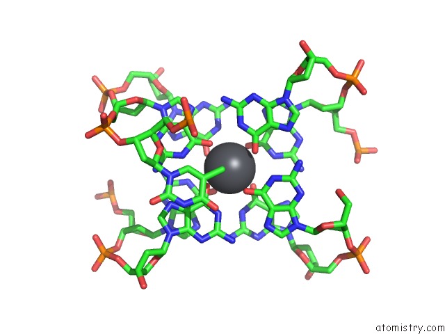
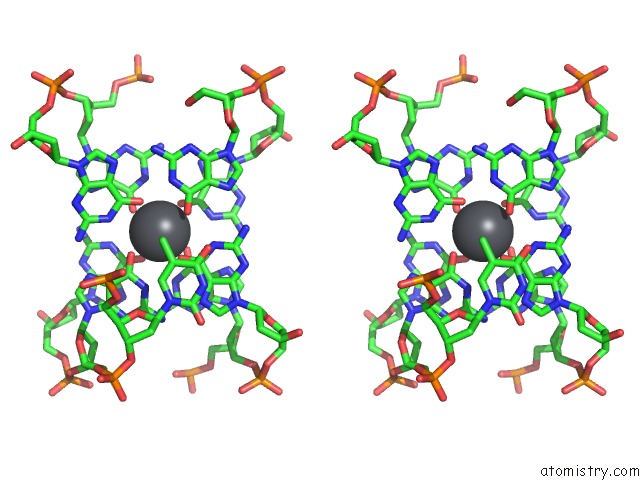
Lead binding site 1 out of 5 in 7d33
Go back to
Lead binding site 1 out
of 5 in the The PB2+ Complexed Structure of Tba G8C Mutant
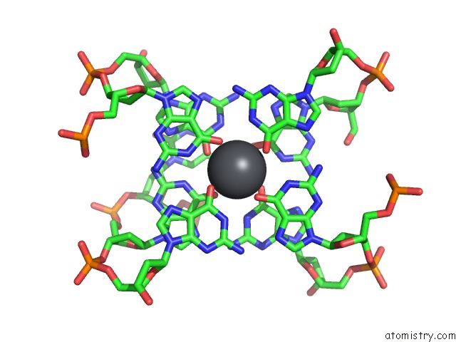
Mono view
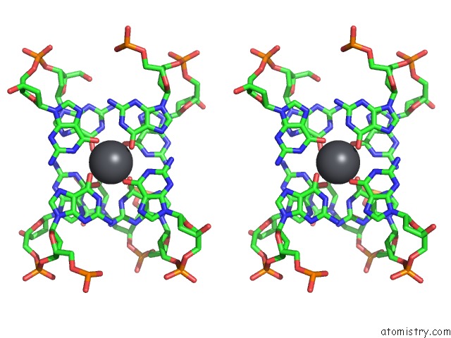
Stereo pair view
Mono view
Stereo pair view
A full contact list of Lead with other atoms in the Pb binding
site number 1 of The PB2+ Complexed Structure of Tba G8C Mutant within 5.0Å range:
|
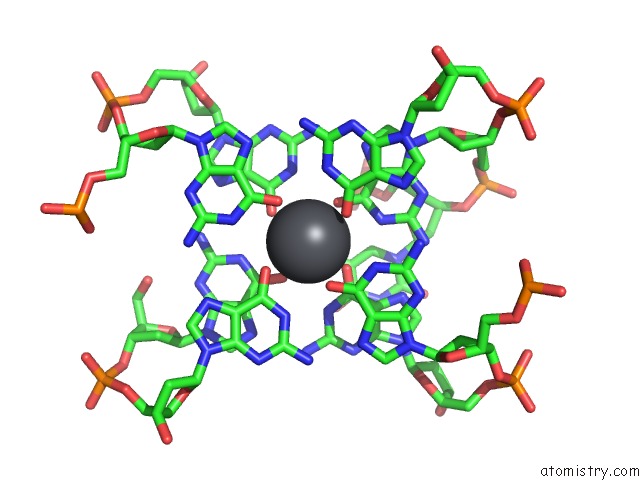
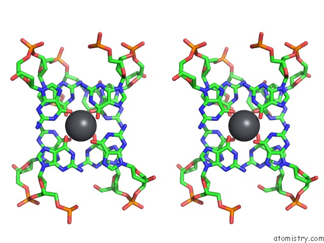
Lead binding site 2 out of 5 in 7d33
Go back to
Lead binding site 2 out
of 5 in the The PB2+ Complexed Structure of Tba G8C Mutant
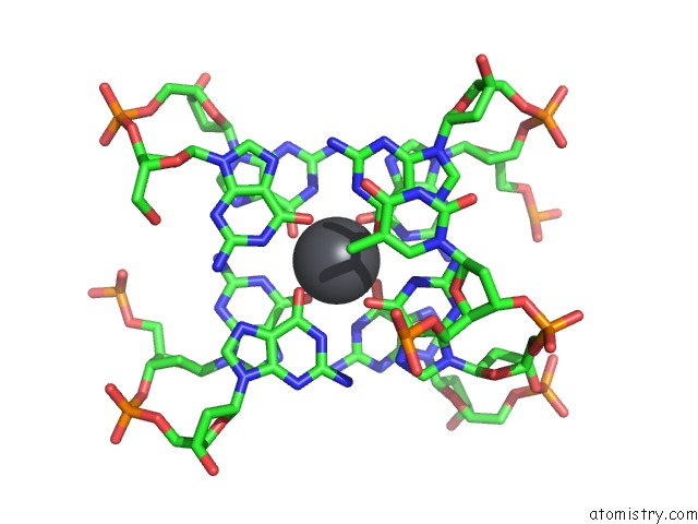
Mono view
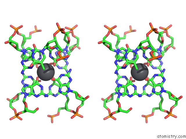
Stereo pair view
Mono view
Stereo pair view
A full contact list of Lead with other atoms in the Pb binding
site number 2 of The PB2+ Complexed Structure of Tba G8C Mutant within 5.0Å range:
|
Lead binding site 3 out of 5 in 7d33
Go back to
Lead binding site 3 out
of 5 in the The PB2+ Complexed Structure of Tba G8C Mutant
Mono view
Stereo pair view
Mono view
Stereo pair view
A full contact list of Lead with other atoms in the Pb binding
site number 3 of The PB2+ Complexed Structure of Tba G8C Mutant within 5.0Å range:
|
Lead binding site 4 out of 5 in 7d33
Go back to
Lead binding site 4 out
of 5 in the The PB2+ Complexed Structure of Tba G8C Mutant
Mono view
Stereo pair view
Mono view
Stereo pair view
A full contact list of Lead with other atoms in the Pb binding
site number 4 of The PB2+ Complexed Structure of Tba G8C Mutant within 5.0Å range:
|
Lead binding site 5 out of 5 in 7d33
Go back to
Lead binding site 5 out
of 5 in the The PB2+ Complexed Structure of Tba G8C Mutant
Mono view
Stereo pair view
Mono view
Stereo pair view
A full contact list of Lead with other atoms in the Pb binding
site number 5 of The PB2+ Complexed Structure of Tba G8C Mutant within 5.0Å range:
|
Reference:
H.H.Liu,
Y.Q.Gao,
J.Sheng,
J.H.Gan.
The PB2+ Complexed Structure of Tba G8C Mutant To Be Published.
Page generated: Mon Aug 18 22:59:08 2025
Last articles
Mn in 9LJUMn in 9LJW
Mn in 9LJS
Mn in 9LJR
Mn in 9LJT
Mn in 9LJV
Mg in 9UA2
Mg in 9R96
Mg in 9VM1
Mg in 9P01