Lead »
PDB 3fhh-8gbm »
4h7x »
Lead in PDB 4h7x: Crystal Structure of the Tetratricopeptide Repeat (Tpr) Motif of Human Dual Specificity Protein Kinase MPS1
Enzymatic activity of Crystal Structure of the Tetratricopeptide Repeat (Tpr) Motif of Human Dual Specificity Protein Kinase MPS1
All present enzymatic activity of Crystal Structure of the Tetratricopeptide Repeat (Tpr) Motif of Human Dual Specificity Protein Kinase MPS1:
2.7.12.1;
2.7.12.1;
Protein crystallography data
The structure of Crystal Structure of the Tetratricopeptide Repeat (Tpr) Motif of Human Dual Specificity Protein Kinase MPS1, PDB code: 4h7x
was solved by
V.M.Bolanos-Garcia,
D.Y.Chirgadze,
T.L.Blundell,
with X-Ray Crystallography technique. A brief refinement statistics is given in the table below:
| Resolution Low / High (Å) | 39.70 / 2.60 |
| Space group | P 41 21 2 |
| Cell size a, b, c (Å), α, β, γ (°) | 79.487, 79.487, 137.684, 90.00, 90.00, 90.00 |
| R / Rfree (%) | 19.3 / 24 |
Lead Binding Sites:
The binding sites of Lead atom in the Crystal Structure of the Tetratricopeptide Repeat (Tpr) Motif of Human Dual Specificity Protein Kinase MPS1
(pdb code 4h7x). This binding sites where shown within
5.0 Angstroms radius around Lead atom.
In total only one binding site of Lead was determined in the Crystal Structure of the Tetratricopeptide Repeat (Tpr) Motif of Human Dual Specificity Protein Kinase MPS1, PDB code: 4h7x:
In total only one binding site of Lead was determined in the Crystal Structure of the Tetratricopeptide Repeat (Tpr) Motif of Human Dual Specificity Protein Kinase MPS1, PDB code: 4h7x:
Lead binding site 1 out of 1 in 4h7x
Go back to
Lead binding site 1 out
of 1 in the Crystal Structure of the Tetratricopeptide Repeat (Tpr) Motif of Human Dual Specificity Protein Kinase MPS1
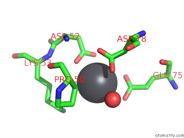
Mono view
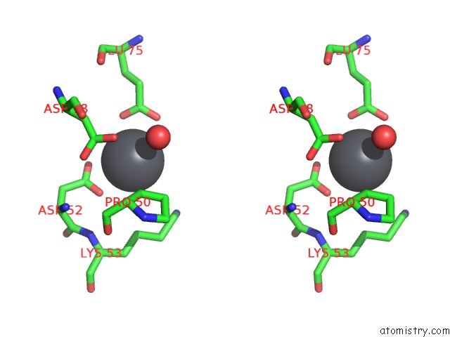
Stereo pair view
Mono view
Stereo pair view
A full contact list of Lead with other atoms in the Pb binding
site number 1 of Crystal Structure of the Tetratricopeptide Repeat (Tpr) Motif of Human Dual Specificity Protein Kinase MPS1 within 5.0Å range:
|
Reference:
P.Thebault,
D.Y.Chirgadze,
Z.Dou,
T.L.Blundell,
S.Elowe,
V.M.Bolanos-Garcia.
Structural and Functional Insights Into the Role of the N-Terminal MPS1 Tpr Domain in the Sac (Spindle Assembly Checkpoint). Biochem.J. V. 448 321 2012.
ISSN: ISSN 0264-6021
PubMed: 23067341
DOI: 10.1042/BJ20121448
Page generated: Mon Aug 18 22:54:39 2025
ISSN: ISSN 0264-6021
PubMed: 23067341
DOI: 10.1042/BJ20121448
Last articles
Mn in 9LJUMn in 9LJW
Mn in 9LJS
Mn in 9LJR
Mn in 9LJT
Mn in 9LJV
Mg in 9UA2
Mg in 9R96
Mg in 9VM1
Mg in 9P01