Lead »
PDB 3fhh-8gbm »
4dl0 »
Lead in PDB 4dl0: Crystal Structure of the Heterotrimeric Egchead Peripheral Stalk Complex of the Yeast Vacuolar Atpase
Enzymatic activity of Crystal Structure of the Heterotrimeric Egchead Peripheral Stalk Complex of the Yeast Vacuolar Atpase
All present enzymatic activity of Crystal Structure of the Heterotrimeric Egchead Peripheral Stalk Complex of the Yeast Vacuolar Atpase:
3.6.3.14;
3.6.3.14;
Protein crystallography data
The structure of Crystal Structure of the Heterotrimeric Egchead Peripheral Stalk Complex of the Yeast Vacuolar Atpase, PDB code: 4dl0
was solved by
R.A.Oot,
L.S.Huang,
E.A.Berry,
S.Wilkens,
with X-Ray Crystallography technique. A brief refinement statistics is given in the table below:
| Resolution Low / High (Å) | 39.71 / 2.91 |
| Space group | P 21 21 21 |
| Cell size a, b, c (Å), α, β, γ (°) | 95.790, 105.611, 120.486, 90.00, 90.00, 90.00 |
| R / Rfree (%) | 20.8 / 25.7 |
Lead Binding Sites:
The binding sites of Lead atom in the Crystal Structure of the Heterotrimeric Egchead Peripheral Stalk Complex of the Yeast Vacuolar Atpase
(pdb code 4dl0). This binding sites where shown within
5.0 Angstroms radius around Lead atom.
In total 2 binding sites of Lead where determined in the Crystal Structure of the Heterotrimeric Egchead Peripheral Stalk Complex of the Yeast Vacuolar Atpase, PDB code: 4dl0:
Jump to Lead binding site number: 1; 2;
In total 2 binding sites of Lead where determined in the Crystal Structure of the Heterotrimeric Egchead Peripheral Stalk Complex of the Yeast Vacuolar Atpase, PDB code: 4dl0:
Jump to Lead binding site number: 1; 2;
Lead binding site 1 out of 2 in 4dl0
Go back to
Lead binding site 1 out
of 2 in the Crystal Structure of the Heterotrimeric Egchead Peripheral Stalk Complex of the Yeast Vacuolar Atpase
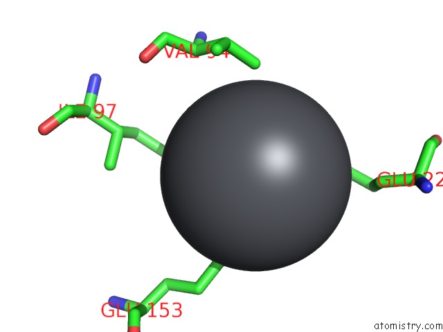
Mono view
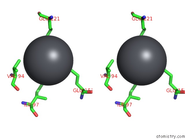
Stereo pair view
Mono view
Stereo pair view
A full contact list of Lead with other atoms in the Pb binding
site number 1 of Crystal Structure of the Heterotrimeric Egchead Peripheral Stalk Complex of the Yeast Vacuolar Atpase within 5.0Å range:
|
Lead binding site 2 out of 2 in 4dl0
Go back to
Lead binding site 2 out
of 2 in the Crystal Structure of the Heterotrimeric Egchead Peripheral Stalk Complex of the Yeast Vacuolar Atpase

Mono view

Stereo pair view
Mono view
Stereo pair view
A full contact list of Lead with other atoms in the Pb binding
site number 2 of Crystal Structure of the Heterotrimeric Egchead Peripheral Stalk Complex of the Yeast Vacuolar Atpase within 5.0Å range:
|
Reference:
R.A.Oot,
L.S.Huang,
E.A.Berry,
S.Wilkens.
Crystal Structure of the Yeast Vacuolar Atpase Heterotrimeric Egc(Head) Peripheral Stalk Complex. Structure V. 20 1881 2012.
ISSN: ISSN 0969-2126
PubMed: 23000382
DOI: 10.1016/J.STR.2012.08.020
Page generated: Mon Aug 18 22:54:38 2025
ISSN: ISSN 0969-2126
PubMed: 23000382
DOI: 10.1016/J.STR.2012.08.020
Last articles
Mn in 9LJUMn in 9LJW
Mn in 9LJS
Mn in 9LJR
Mn in 9LJT
Mn in 9LJV
Mg in 9UA2
Mg in 9R96
Mg in 9VM1
Mg in 9P01