Lead »
PDB 1afv-3ec8 »
2o3c »
Lead in PDB 2o3c: Crystal Structure of Zebrafish Ape
Protein crystallography data
The structure of Crystal Structure of Zebrafish Ape, PDB code: 2o3c
was solved by
M.M.Georgiadis,
R.K.Gaur,
S.Delaplane,
J.Svenson,
with X-Ray Crystallography technique. A brief refinement statistics is given in the table below:
| Resolution Low / High (Å) | 50.00 / 2.30 |
| Space group | P 1 21 1 |
| Cell size a, b, c (Å), α, β, γ (°) | 54.660, 117.780, 85.840, 90.00, 98.34, 90.00 |
| R / Rfree (%) | 19.9 / 23.6 |
Lead Binding Sites:
The binding sites of Lead atom in the Crystal Structure of Zebrafish Ape
(pdb code 2o3c). This binding sites where shown within
5.0 Angstroms radius around Lead atom.
In total 3 binding sites of Lead where determined in the Crystal Structure of Zebrafish Ape, PDB code: 2o3c:
Jump to Lead binding site number: 1; 2; 3;
In total 3 binding sites of Lead where determined in the Crystal Structure of Zebrafish Ape, PDB code: 2o3c:
Jump to Lead binding site number: 1; 2; 3;
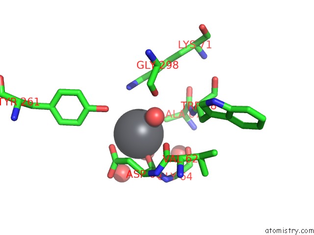
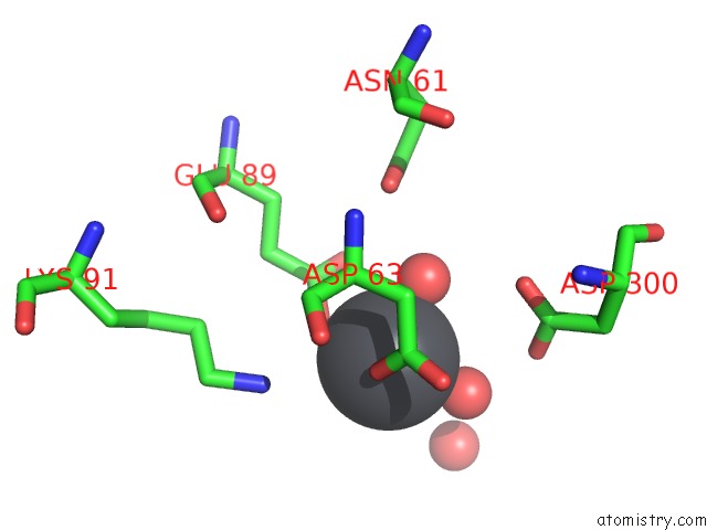
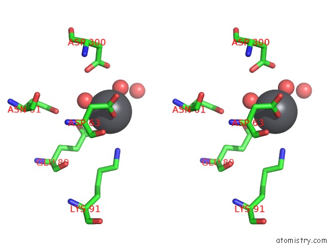
Lead binding site 1 out of 3 in 2o3c
Go back to
Lead binding site 1 out
of 3 in the Crystal Structure of Zebrafish Ape
Mono view
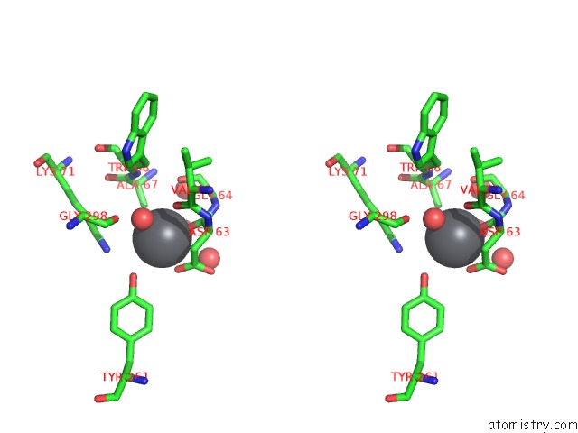
Stereo pair view
Mono view
Stereo pair view
A full contact list of Lead with other atoms in the Pb binding
site number 1 of Crystal Structure of Zebrafish Ape within 5.0Å range:
|
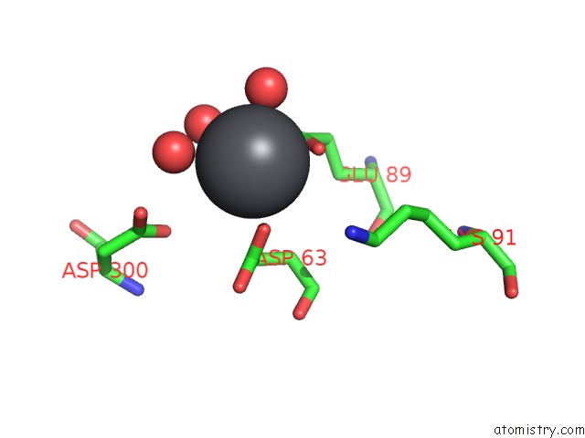
Lead binding site 2 out of 3 in 2o3c
Go back to
Lead binding site 2 out
of 3 in the Crystal Structure of Zebrafish Ape
Mono view
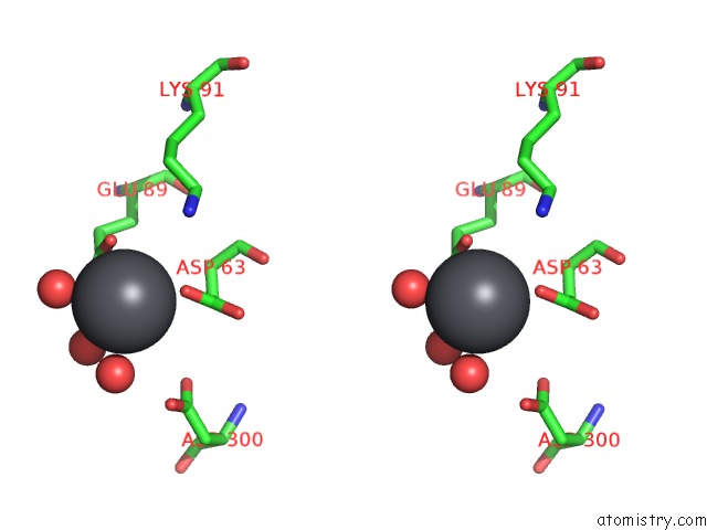
Stereo pair view
Mono view
Stereo pair view
A full contact list of Lead with other atoms in the Pb binding
site number 2 of Crystal Structure of Zebrafish Ape within 5.0Å range:
|
Lead binding site 3 out of 3 in 2o3c
Go back to
Lead binding site 3 out
of 3 in the Crystal Structure of Zebrafish Ape
Mono view
Stereo pair view
Mono view
Stereo pair view
A full contact list of Lead with other atoms in the Pb binding
site number 3 of Crystal Structure of Zebrafish Ape within 5.0Å range:
|
Reference:
M.M.Georgiadis,
M.Luo,
R.K.Gaur,
S.Delaplane,
X.Li,
M.R.Kelley.
Evolution of the Redox Function in Mammalian Apurinic/Apyrimidinic Endonuclease Mutat.Res. V. 643 54 2008.
ISSN: ISSN 0027-5107
PubMed: 18579163
DOI: 10.1016/J.MRFMMM.2008.04.008
Page generated: Thu Oct 10 10:04:54 2024
ISSN: ISSN 0027-5107
PubMed: 18579163
DOI: 10.1016/J.MRFMMM.2008.04.008
Last articles
Br in 1C4UBr in 1BSO
Br in 1BK9
Br in 1AZF
Br in 1AWC
Br in 1A35
Br in 1AQV
Br in 1AIO
Br in 1A3E
Br in 115D