Lead »
PDB 1afv-3ec8 »
1xxa »
Lead in PDB 1xxa: C-Terminal Domain of Escherichia Coli Arginine Repressor/ L- Arginine Complex; Pb Derivative
Protein crystallography data
The structure of C-Terminal Domain of Escherichia Coli Arginine Repressor/ L- Arginine Complex; Pb Derivative, PDB code: 1xxa
was solved by
G.D.Van Duyne,
G.Ghosh,
W.K.Maas,
P.B.Sigler,
with X-Ray Crystallography technique. A brief refinement statistics is given in the table below:
| Resolution Low / High (Å) | 8.00 / 2.20 |
| Space group | C 2 2 21 |
| Cell size a, b, c (Å), α, β, γ (°) | 53.500, 83.800, 217.000, 90.00, 90.00, 90.00 |
| R / Rfree (%) | 20 / 33 |
Lead Binding Sites:
The binding sites of Lead atom in the C-Terminal Domain of Escherichia Coli Arginine Repressor/ L- Arginine Complex; Pb Derivative
(pdb code 1xxa). This binding sites where shown within
5.0 Angstroms radius around Lead atom.
In total 4 binding sites of Lead where determined in the C-Terminal Domain of Escherichia Coli Arginine Repressor/ L- Arginine Complex; Pb Derivative, PDB code: 1xxa:
Jump to Lead binding site number: 1; 2; 3; 4;
In total 4 binding sites of Lead where determined in the C-Terminal Domain of Escherichia Coli Arginine Repressor/ L- Arginine Complex; Pb Derivative, PDB code: 1xxa:
Jump to Lead binding site number: 1; 2; 3; 4;
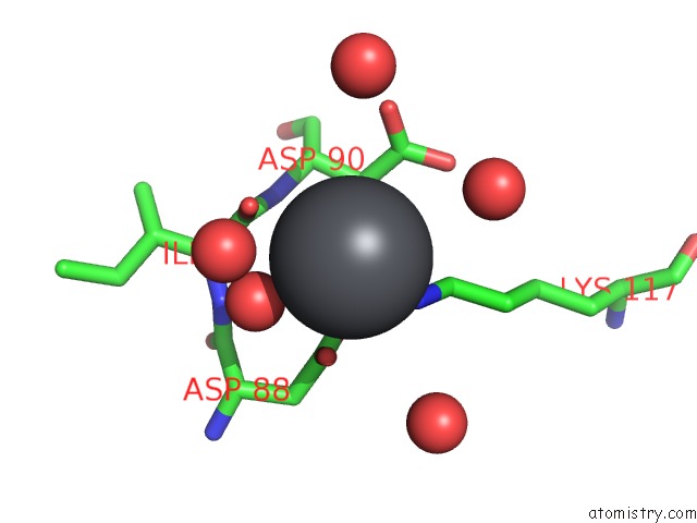
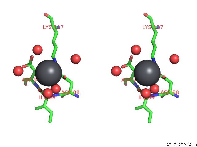
Lead binding site 1 out of 4 in 1xxa
Go back to
Lead binding site 1 out
of 4 in the C-Terminal Domain of Escherichia Coli Arginine Repressor/ L- Arginine Complex; Pb Derivative
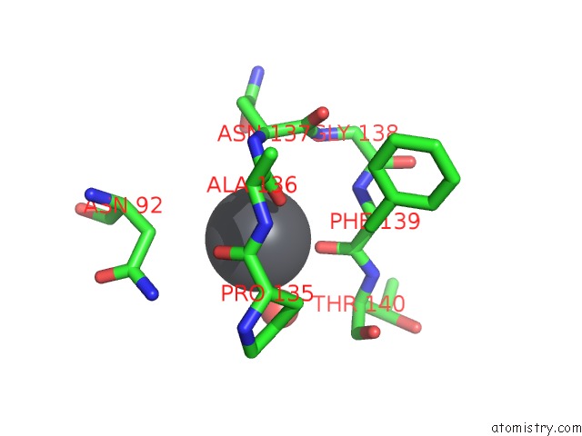
Mono view
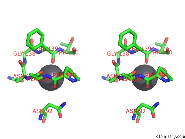
Stereo pair view
Mono view
Stereo pair view
A full contact list of Lead with other atoms in the Pb binding
site number 1 of C-Terminal Domain of Escherichia Coli Arginine Repressor/ L- Arginine Complex; Pb Derivative within 5.0Å range:
|
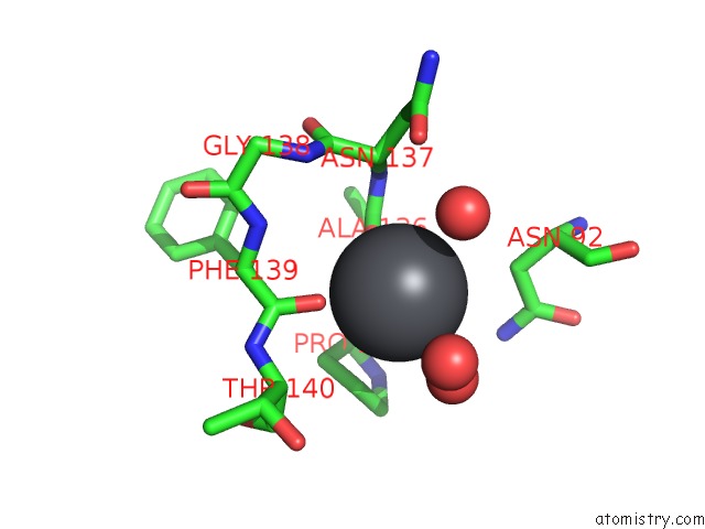
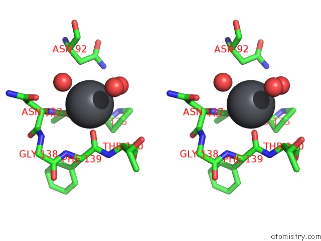
Lead binding site 2 out of 4 in 1xxa
Go back to
Lead binding site 2 out
of 4 in the C-Terminal Domain of Escherichia Coli Arginine Repressor/ L- Arginine Complex; Pb Derivative
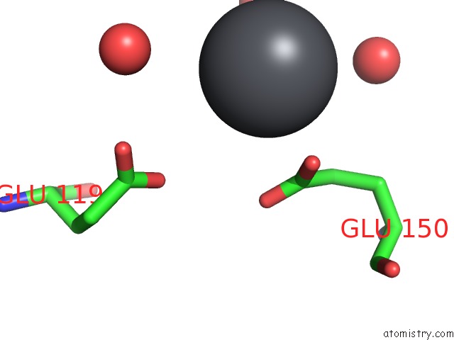
Mono view
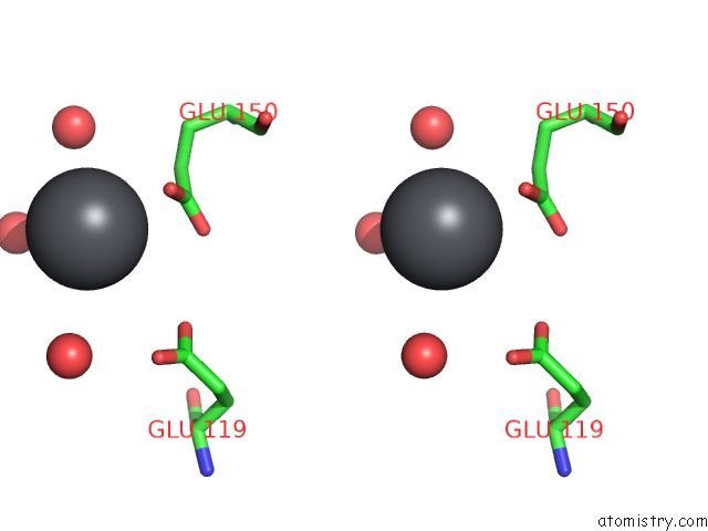
Stereo pair view
Mono view
Stereo pair view
A full contact list of Lead with other atoms in the Pb binding
site number 2 of C-Terminal Domain of Escherichia Coli Arginine Repressor/ L- Arginine Complex; Pb Derivative within 5.0Å range:
|
Lead binding site 3 out of 4 in 1xxa
Go back to
Lead binding site 3 out
of 4 in the C-Terminal Domain of Escherichia Coli Arginine Repressor/ L- Arginine Complex; Pb Derivative
Mono view
Stereo pair view
Mono view
Stereo pair view
A full contact list of Lead with other atoms in the Pb binding
site number 3 of C-Terminal Domain of Escherichia Coli Arginine Repressor/ L- Arginine Complex; Pb Derivative within 5.0Å range:
|
Lead binding site 4 out of 4 in 1xxa
Go back to
Lead binding site 4 out
of 4 in the C-Terminal Domain of Escherichia Coli Arginine Repressor/ L- Arginine Complex; Pb Derivative
Mono view
Stereo pair view
Mono view
Stereo pair view
A full contact list of Lead with other atoms in the Pb binding
site number 4 of C-Terminal Domain of Escherichia Coli Arginine Repressor/ L- Arginine Complex; Pb Derivative within 5.0Å range:
|
Reference:
G.D.Van Duyne,
G.Ghosh,
W.K.Maas,
P.B.Sigler.
Structure of the Oligomerization and L-Arginine Binding Domain of the Arginine Repressor of Escherichia Coli. J.Mol.Biol. V. 256 377 1996.
ISSN: ISSN 0022-2836
PubMed: 8594204
DOI: 10.1006/JMBI.1996.0093
Page generated: Thu Oct 10 10:02:55 2024
ISSN: ISSN 0022-2836
PubMed: 8594204
DOI: 10.1006/JMBI.1996.0093
Last articles
Zn in 9MJ5Zn in 9HNW
Zn in 9G0L
Zn in 9FNE
Zn in 9DZN
Zn in 9E0I
Zn in 9D32
Zn in 9DAK
Zn in 8ZXC
Zn in 8ZUF