Lead »
PDB 1afv-3ec8 »
1sn8 »
Lead in PDB 1sn8: Crystal Structure of the S1 Domain of Rnase E From E. Coli (Pb Derivative)
Protein crystallography data
The structure of Crystal Structure of the S1 Domain of Rnase E From E. Coli (Pb Derivative), PDB code: 1sn8
was solved by
M.Schubert,
R.E.Edge,
P.Lario,
M.A.Cook,
N.C.J.Strynadka,
G.A.Mackie,
L.P.Mcintosh,
with X-Ray Crystallography technique. A brief refinement statistics is given in the table below:
| Resolution Low / High (Å) | 25.00 / 2.00 |
| Space group | P 41 21 2 |
| Cell size a, b, c (Å), α, β, γ (°) | 70.580, 70.580, 87.902, 90.00, 90.00, 90.00 |
| R / Rfree (%) | 18.2 / 23.2 |
Lead Binding Sites:
The binding sites of Lead atom in the Crystal Structure of the S1 Domain of Rnase E From E. Coli (Pb Derivative)
(pdb code 1sn8). This binding sites where shown within
5.0 Angstroms radius around Lead atom.
In total 2 binding sites of Lead where determined in the Crystal Structure of the S1 Domain of Rnase E From E. Coli (Pb Derivative), PDB code: 1sn8:
Jump to Lead binding site number: 1; 2;
In total 2 binding sites of Lead where determined in the Crystal Structure of the S1 Domain of Rnase E From E. Coli (Pb Derivative), PDB code: 1sn8:
Jump to Lead binding site number: 1; 2;
Lead binding site 1 out of 2 in 1sn8
Go back to
Lead binding site 1 out
of 2 in the Crystal Structure of the S1 Domain of Rnase E From E. Coli (Pb Derivative)
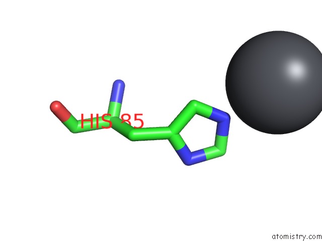
Mono view
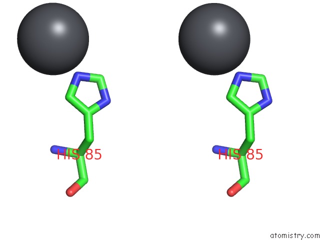
Stereo pair view
Mono view
Stereo pair view
A full contact list of Lead with other atoms in the Pb binding
site number 1 of Crystal Structure of the S1 Domain of Rnase E From E. Coli (Pb Derivative) within 5.0Å range:
|
Lead binding site 2 out of 2 in 1sn8
Go back to
Lead binding site 2 out
of 2 in the Crystal Structure of the S1 Domain of Rnase E From E. Coli (Pb Derivative)
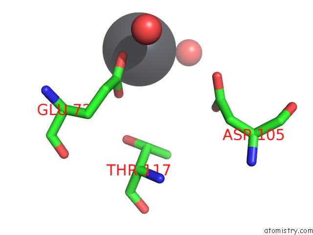
Mono view
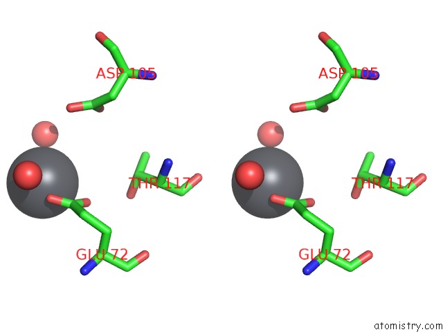
Stereo pair view
Mono view
Stereo pair view
A full contact list of Lead with other atoms in the Pb binding
site number 2 of Crystal Structure of the S1 Domain of Rnase E From E. Coli (Pb Derivative) within 5.0Å range:
|
Reference:
M.Schubert,
R.E.Edge,
P.Lario,
M.A.Cook,
N.C.Strynadka,
G.A.Mackie,
L.P.Mcintosh.
Structural Characterization of the Rnase E S1 Domain and Identification of Its Oligonucleotide-Binding and Dimerization Interfaces. J.Mol.Biol. V. 341 37 2004.
ISSN: ISSN 0022-2836
PubMed: 15312761
DOI: 10.1016/J.JMB.2004.05.061
Page generated: Thu Oct 10 10:02:13 2024
ISSN: ISSN 0022-2836
PubMed: 15312761
DOI: 10.1016/J.JMB.2004.05.061
Last articles
Zn in 9J0NZn in 9J0O
Zn in 9J0P
Zn in 9FJX
Zn in 9EKB
Zn in 9C0F
Zn in 9CAH
Zn in 9CH0
Zn in 9CH3
Zn in 9CH1