Lead »
PDB 1afv-3ec8 »
1qr7 »
Lead in PDB 1qr7: Crystal Structure of Phenylalanine-Regulated 3-Deoxy-D- Arabino-Heptulosonate-7-Phosphate Synthase From Escherichia Coli Complexed with PB2+ and Pep
Enzymatic activity of Crystal Structure of Phenylalanine-Regulated 3-Deoxy-D- Arabino-Heptulosonate-7-Phosphate Synthase From Escherichia Coli Complexed with PB2+ and Pep
All present enzymatic activity of Crystal Structure of Phenylalanine-Regulated 3-Deoxy-D- Arabino-Heptulosonate-7-Phosphate Synthase From Escherichia Coli Complexed with PB2+ and Pep:
4.1.2.15;
4.1.2.15;
Protein crystallography data
The structure of Crystal Structure of Phenylalanine-Regulated 3-Deoxy-D- Arabino-Heptulosonate-7-Phosphate Synthase From Escherichia Coli Complexed with PB2+ and Pep, PDB code: 1qr7
was solved by
I.A.Shumilin,
R.H.Kretsinger,
R.H.Bauerle,
with X-Ray Crystallography technique. A brief refinement statistics is given in the table below:
| Resolution Low / High (Å) | 18.00 / 2.60 |
| Space group | C 1 2 1 |
| Cell size a, b, c (Å), α, β, γ (°) | 211.731, 51.326, 148.099, 90.00, 116.43, 90.00 |
| R / Rfree (%) | 19.4 / 25.6 |
Lead Binding Sites:
The binding sites of Lead atom in the Crystal Structure of Phenylalanine-Regulated 3-Deoxy-D- Arabino-Heptulosonate-7-Phosphate Synthase From Escherichia Coli Complexed with PB2+ and Pep
(pdb code 1qr7). This binding sites where shown within
5.0 Angstroms radius around Lead atom.
In total 4 binding sites of Lead where determined in the Crystal Structure of Phenylalanine-Regulated 3-Deoxy-D- Arabino-Heptulosonate-7-Phosphate Synthase From Escherichia Coli Complexed with PB2+ and Pep, PDB code: 1qr7:
Jump to Lead binding site number: 1; 2; 3; 4;
In total 4 binding sites of Lead where determined in the Crystal Structure of Phenylalanine-Regulated 3-Deoxy-D- Arabino-Heptulosonate-7-Phosphate Synthase From Escherichia Coli Complexed with PB2+ and Pep, PDB code: 1qr7:
Jump to Lead binding site number: 1; 2; 3; 4;
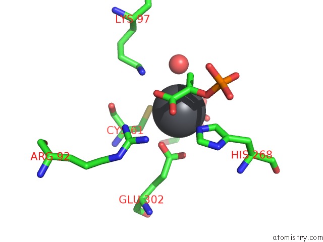
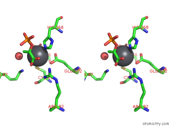
Lead binding site 1 out of 4 in 1qr7
Go back to
Lead binding site 1 out
of 4 in the Crystal Structure of Phenylalanine-Regulated 3-Deoxy-D- Arabino-Heptulosonate-7-Phosphate Synthase From Escherichia Coli Complexed with PB2+ and Pep
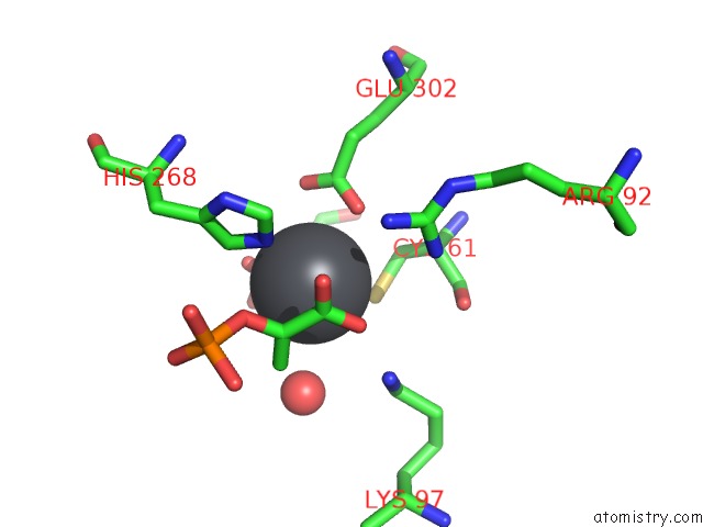
Mono view
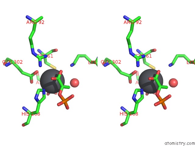
Stereo pair view
Mono view
Stereo pair view
A full contact list of Lead with other atoms in the Pb binding
site number 1 of Crystal Structure of Phenylalanine-Regulated 3-Deoxy-D- Arabino-Heptulosonate-7-Phosphate Synthase From Escherichia Coli Complexed with PB2+ and Pep within 5.0Å range:
|
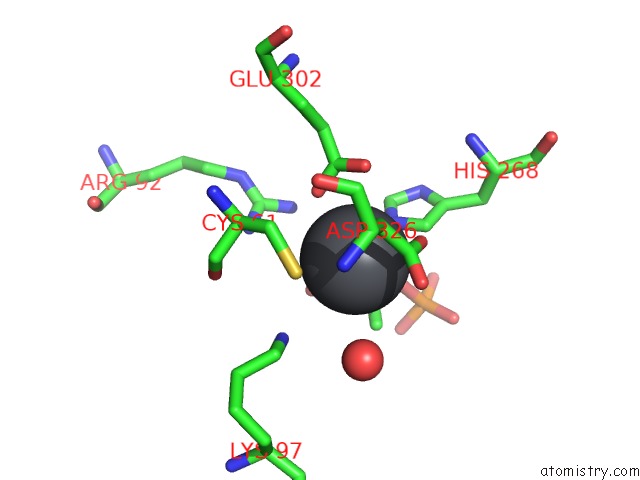
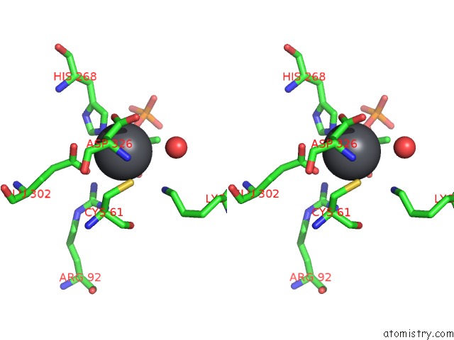
Lead binding site 2 out of 4 in 1qr7
Go back to
Lead binding site 2 out
of 4 in the Crystal Structure of Phenylalanine-Regulated 3-Deoxy-D- Arabino-Heptulosonate-7-Phosphate Synthase From Escherichia Coli Complexed with PB2+ and Pep
Mono view
Stereo pair view
Mono view
Stereo pair view
A full contact list of Lead with other atoms in the Pb binding
site number 2 of Crystal Structure of Phenylalanine-Regulated 3-Deoxy-D- Arabino-Heptulosonate-7-Phosphate Synthase From Escherichia Coli Complexed with PB2+ and Pep within 5.0Å range:
|
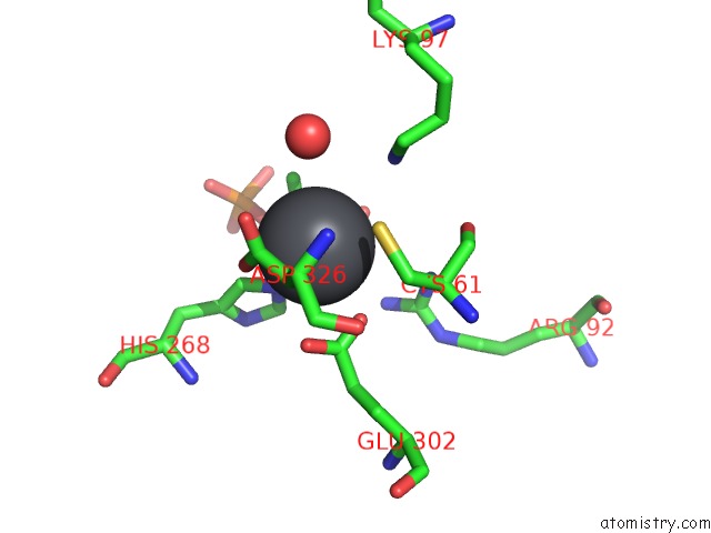
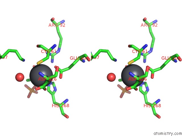
Lead binding site 3 out of 4 in 1qr7
Go back to
Lead binding site 3 out
of 4 in the Crystal Structure of Phenylalanine-Regulated 3-Deoxy-D- Arabino-Heptulosonate-7-Phosphate Synthase From Escherichia Coli Complexed with PB2+ and Pep
Mono view
Stereo pair view
Mono view
Stereo pair view
A full contact list of Lead with other atoms in the Pb binding
site number 3 of Crystal Structure of Phenylalanine-Regulated 3-Deoxy-D- Arabino-Heptulosonate-7-Phosphate Synthase From Escherichia Coli Complexed with PB2+ and Pep within 5.0Å range:
|
Lead binding site 4 out of 4 in 1qr7
Go back to
Lead binding site 4 out
of 4 in the Crystal Structure of Phenylalanine-Regulated 3-Deoxy-D- Arabino-Heptulosonate-7-Phosphate Synthase From Escherichia Coli Complexed with PB2+ and Pep
Mono view
Stereo pair view
Mono view
Stereo pair view
A full contact list of Lead with other atoms in the Pb binding
site number 4 of Crystal Structure of Phenylalanine-Regulated 3-Deoxy-D- Arabino-Heptulosonate-7-Phosphate Synthase From Escherichia Coli Complexed with PB2+ and Pep within 5.0Å range:
|
Reference:
I.A.Shumilin,
R.H.Kretsinger,
R.H.Bauerle.
Crystal Structure of Phenylalanine-Regulated 3-Deoxy-D-Arabino-Heptulosonate-7-Phosphate Synthase From Escherichia Coli. Structure Fold.Des. V. 7 865 1999.
ISSN: ISSN 0969-2126
PubMed: 10425687
DOI: 10.1016/S0969-2126(99)80109-9
Page generated: Thu Oct 10 10:01:59 2024
ISSN: ISSN 0969-2126
PubMed: 10425687
DOI: 10.1016/S0969-2126(99)80109-9
Last articles
Zn in 9J0NZn in 9J0O
Zn in 9J0P
Zn in 9FJX
Zn in 9EKB
Zn in 9C0F
Zn in 9CAH
Zn in 9CH0
Zn in 9CH3
Zn in 9CH1