Atomistry » Lead » PDB 1afv-3ec8 » 1n0y
Atomistry »
  Lead »
    PDB 1afv-3ec8 »
      1n0y »

Lead in PDB 1n0y: Crystal Structure of Pb-Bound Calmodulin

Protein crystallography data

The structure of Crystal Structure of Pb-Bound Calmodulin, PDB code: 1n0y was solved by M.A.Wilson, A.T.Brunger, with X-Ray Crystallography technique. A brief refinement statistics is given in the table below:

Resolution Low / High (Å) 53.00 / 1.75
Space group C 1 2 1
Cell size a, b, c (Å), α, β, γ (°) 100.936, 30.792, 113.084, 90.00, 109.31, 90.00
R / Rfree (%) 21.3 / 22.3

Other elements in 1n0y:

The structure of Crystal Structure of Pb-Bound Calmodulin also contains other interesting chemical elements:

Arsenic (As) 1 atom

Lead Binding Sites:

Pages:

>>> Page 1 <<< Page 2, Binding sites: 11 - 14;

Binding sites:

The binding sites of Lead atom in the Crystal Structure of Pb-Bound Calmodulin (pdb code 1n0y). This binding sites where shown within 5.0 Angstroms radius around Lead atom.
In total 14 binding sites of Lead where determined in the Crystal Structure of Pb-Bound Calmodulin, PDB code: 1n0y:
Jump to Lead binding site number: 1; 2; 3; 4; 5; 6; 7; 8; 9; 10;

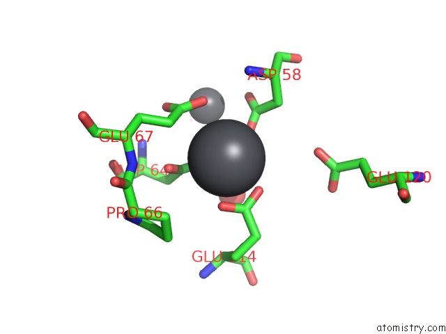
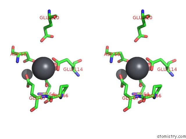
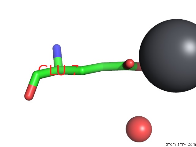
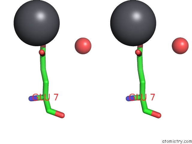
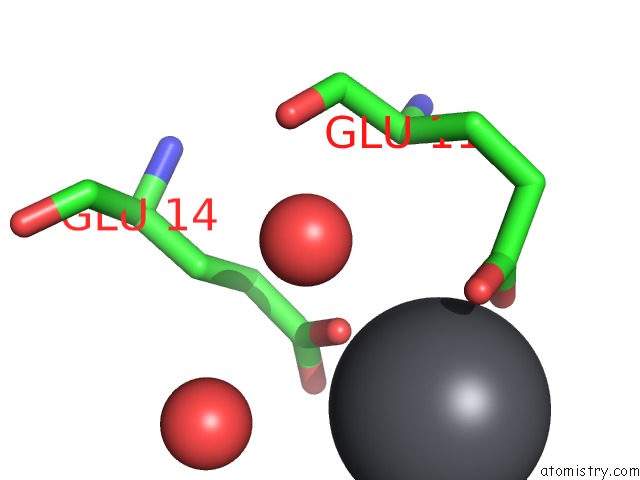
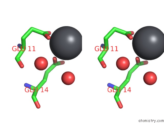
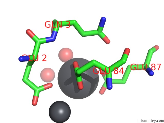
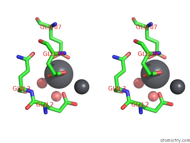
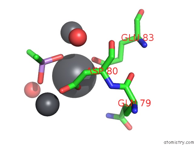
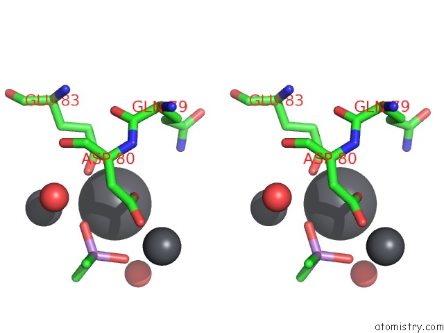
Lead binding site 1 out of 14 in 1n0y

Go back to Lead Binding Sites List in 1n0y
Lead binding site 1 out of 14 in the Crystal Structure of Pb-Bound Calmodulin


Mono view


Stereo pair view

A full contact list of Lead with other atoms in the Pb binding site number 1 of Crystal Structure of Pb-Bound Calmodulin within 5.0Å range:
probe atom residue distance (Å) B Occ
A:Pb500

b:17.0
occ:1.00
OD2 A:ASP64 2.4 17.1 1.0
OD2 A:ASP58 2.4 19.2 1.0
OE1 B:GLU114 2.5 20.8 1.0
OE2 B:GLU114 2.5 18.1 1.0
CD B:GLU114 2.8 19.6 1.0
CG A:ASP64 3.1 16.8 1.0
OD1 A:ASP64 3.2 14.1 1.0
OE2 A:GLU67 3.2 21.2 1.0
CG A:ASP58 3.4 21.2 1.0
CD A:GLU67 3.5 16.7 1.0
CG A:GLU67 3.6 17.7 1.0
CB A:ASP58 4.0 23.7 1.0
O B:HOH2004 4.3 14.3 1.0
CG B:GLU114 4.4 16.6 1.0
OD1 A:ASP58 4.4 19.2 1.0
OE1 A:GLU67 4.4 15.5 1.0
PB A:PB501 4.4 19.0 1.0
CB A:ASP64 4.5 14.4 1.0
N A:ASP58 4.7 24.5 1.0
CB A:PRO66 4.7 15.1 1.0
OE2 B:GLU120 4.9 30.5 1.0
CB A:GLU67 5.0 15.6 1.0

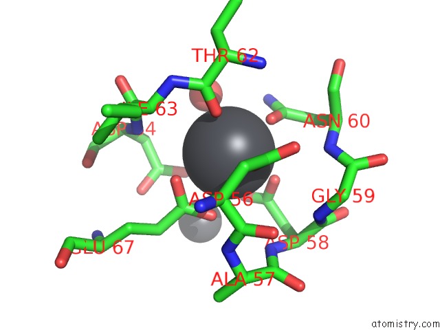
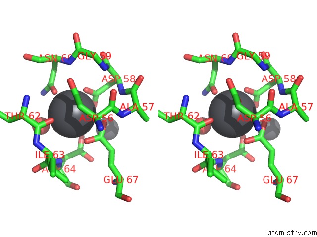
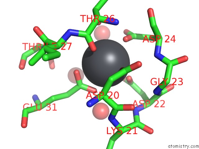
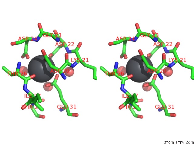
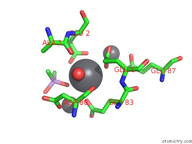
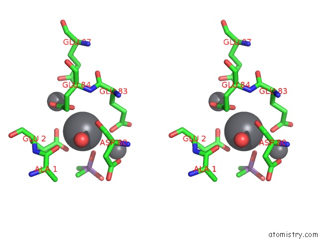
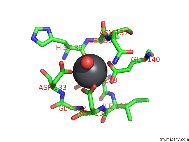
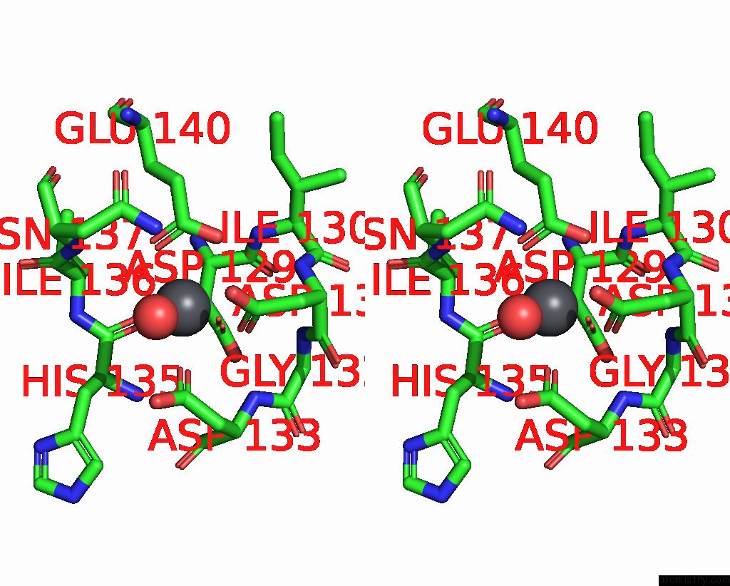
Lead binding site 2 out of 14 in 1n0y

Go back to Lead Binding Sites List in 1n0y
Lead binding site 2 out of 14 in the Crystal Structure of Pb-Bound Calmodulin


Mono view


Stereo pair view

A full contact list of Lead with other atoms in the Pb binding site number 2 of Crystal Structure of Pb-Bound Calmodulin within 5.0Å range:
probe atom residue distance (Å) B Occ
A:Pb501

b:19.0
occ:1.00
OD1 A:ASP56 2.2 25.1 1.0
OD1 A:ASN60 2.4 23.7 1.0
OD1 A:ASP58 2.5 19.2 1.0
O A:THR62 2.5 19.1 1.0
OE1 A:GLU67 2.8 15.5 1.0
OE2 A:GLU67 2.8 21.2 1.0
CD A:GLU67 3.1 16.7 1.0
CG A:ASP58 3.1 21.2 1.0
OD2 A:ASP58 3.1 19.2 1.0
CG A:ASN60 3.1 27.3 1.0
O A:HOH1015 3.2 16.4 1.0
OD2 A:ASP64 3.3 17.1 1.0
CG A:ASP56 3.4 26.6 1.0
ND2 A:ASN60 3.6 28.4 1.0
C A:THR62 3.7 19.0 1.0
CG A:ASP64 4.0 16.8 1.0
N A:ASP64 4.1 13.4 1.0
OD2 A:ASP56 4.2 30.2 1.0
CB A:ASN60 4.3 29.1 1.0
CA A:ILE63 4.3 15.9 1.0
CB A:ASP56 4.4 26.9 1.0
PB A:PB500 4.4 17.0 1.0
N A:ASN60 4.4 31.3 1.0
N A:ILE63 4.4 16.2 1.0
CA A:ASP56 4.5 24.9 1.0
CB A:ASP58 4.5 23.7 1.0
CG A:GLU67 4.5 17.7 1.0
N A:THR62 4.5 23.5 1.0
N A:ASP58 4.5 24.5 1.0
C A:ILE63 4.6 14.6 1.0
OG1 A:THR62 4.6 18.6 1.0
OD1 A:ASP64 4.6 14.1 1.0
CB A:ASP64 4.6 14.4 1.0
CA A:THR62 4.7 21.1 1.0
C A:ASP56 4.8 26.0 1.0
N A:GLY59 4.9 29.3 1.0
CA A:ASN60 4.9 31.7 1.0
N A:ALA57 4.9 24.3 1.0
CA A:ASP58 4.9 25.8 1.0
C A:ASP58 5.0 28.3 1.0
CA A:ASP64 5.0 13.4 1.0

Lead binding site 3 out of 14 in 1n0y

Go back to Lead Binding Sites List in 1n0y
Lead binding site 3 out of 14 in the Crystal Structure of Pb-Bound Calmodulin


Mono view


Stereo pair view

A full contact list of Lead with other atoms in the Pb binding site number 3 of Crystal Structure of Pb-Bound Calmodulin within 5.0Å range:
probe atom residue distance (Å) B Occ
A:Pb502

b:20.8
occ:1.00
OE2 A:GLU7 2.4 27.2 1.0
OE1 A:GLU7 2.6 21.0 1.0
CD A:GLU7 2.8 19.8 1.0
CG A:GLU7 4.3 17.5 1.0
O A:HOH1011 4.8 28.8 1.0

Lead binding site 4 out of 14 in 1n0y

Go back to Lead Binding Sites List in 1n0y
Lead binding site 4 out of 14 in the Crystal Structure of Pb-Bound Calmodulin


Mono view


Stereo pair view

A full contact list of Lead with other atoms in the Pb binding site number 4 of Crystal Structure of Pb-Bound Calmodulin within 5.0Å range:
probe atom residue distance (Å) B Occ
A:Pb503

b:18.9
occ:1.00
OD1 A:ASP20 2.3 15.5 1.0
OD1 A:ASP22 2.5 15.9 1.0
OE2 A:GLU31 2.5 17.6 1.0
OE1 A:GLU31 2.6 19.3 1.0
O A:HOH1006 2.6 28.9 1.0
OD1 A:ASP24 2.6 18.2 1.0
O A:THR26 2.7 15.9 1.0
CD A:GLU31 2.9 17.4 1.0
CG A:ASP24 3.4 19.3 1.0
CG A:ASP20 3.4 15.2 1.0
CG A:ASP22 3.4 21.9 1.0
OG1 A:THR26 3.5 17.9 1.0
C A:THR26 3.6 14.8 1.0
OD2 A:ASP22 3.7 26.5 1.0
OD2 A:ASP24 3.8 25.1 1.0
CA A:ASP20 4.0 14.2 1.0
CB A:ASP20 4.1 13.1 1.0
N A:THR26 4.3 14.0 1.0
CG A:GLU31 4.3 22.5 1.0
N A:ASP24 4.4 17.1 1.0
OD2 A:ASP20 4.4 15.8 1.0
CA A:THR26 4.4 14.1 1.0
O A:HOH1026 4.5 41.3 1.0
CB A:ASP24 4.5 19.6 1.0
C A:ASP20 4.5 15.4 1.0
CB A:THR26 4.5 15.1 1.0
N A:ILE27 4.5 14.9 1.0
O A:HOH1013 4.7 22.2 1.0
N A:ASP22 4.7 19.8 1.0
O A:HOH1063 4.7 31.1 1.0
CG2 A:THR28 4.7 24.4 1.0
CA A:ILE27 4.7 16.2 1.0
N A:LYS21 4.8 16.9 1.0
CB A:ASP22 4.8 22.0 1.0
N A:GLY23 4.9 17.4 1.0
CA A:ASP24 4.9 17.5 1.0
N A:THR28 4.9 19.7 1.0

Lead binding site 5 out of 14 in 1n0y

Go back to Lead Binding Sites List in 1n0y
Lead binding site 5 out of 14 in the Crystal Structure of Pb-Bound Calmodulin


Mono view


Stereo pair view

A full contact list of Lead with other atoms in the Pb binding site number 5 of Crystal Structure of Pb-Bound Calmodulin within 5.0Å range:
probe atom residue distance (Å) B Occ
A:Pb504

b:32.0
occ:1.00
OE1 A:GLU14 2.3 30.8 1.0
OE2 A:GLU11 2.5 23.3 1.0
OE1 A:GLU11 2.7 22.3 1.0
CD A:GLU11 2.9 21.5 1.0
CD A:GLU14 3.0 30.4 1.0
OE2 A:GLU14 3.0 36.7 1.0
CG A:GLU11 4.4 16.7 1.0
CG A:GLU14 4.5 23.8 1.0
O A:HOH1070 4.7 45.7 1.0
O A:HOH1024 4.7 28.7 1.0

Lead binding site 6 out of 14 in 1n0y

Go back to Lead Binding Sites List in 1n0y
Lead binding site 6 out of 14 in the Crystal Structure of Pb-Bound Calmodulin


Mono view


Stereo pair view

A full contact list of Lead with other atoms in the Pb binding site number 6 of Crystal Structure of Pb-Bound Calmodulin within 5.0Å range:
probe atom residue distance (Å) B Occ
A:Pb507

b:21.2
occ:1.00
O2 A:CAC1001 2.5 24.1 1.0
OE1 A:GLU2 2.5 37.7 1.0
OE2 A:GLU2 2.7 34.4 1.0
O A:HOH1020 2.9 22.5 1.0
CD A:GLU2 2.9 35.8 1.0
OE2 B:GLU84 3.0 18.2 1.0
OE1 B:GLU83 3.1 25.2 1.0
AS A:CAC1001 3.8 22.7 1.0
CD B:GLU84 3.9 17.9 1.0
PB B:PB506 4.0 22.0 1.0
PB B:PB505 4.1 19.3 1.0
CD B:GLU83 4.1 25.2 1.0
C1 A:CAC1001 4.2 25.6 1.0
CG A:GLU2 4.4 34.8 1.0
CB B:GLU83 4.6 21.9 1.0
C2 A:CAC1001 4.6 24.6 1.0
OE1 B:GLU84 4.6 19.6 1.0
CG B:GLU84 4.6 19.4 1.0
OE2 B:GLU83 4.8 27.9 1.0
O A:ALA1 4.8 47.2 1.0
OE1 B:GLU87 4.8 22.8 1.0
O B:ASP80 4.9 21.2 1.0
CG B:GLU83 5.0 24.6 1.0

Lead binding site 7 out of 14 in 1n0y

Go back to Lead Binding Sites List in 1n0y
Lead binding site 7 out of 14 in the Crystal Structure of Pb-Bound Calmodulin


Mono view


Stereo pair view

A full contact list of Lead with other atoms in the Pb binding site number 7 of Crystal Structure of Pb-Bound Calmodulin within 5.0Å range:

Lead binding site 8 out of 14 in 1n0y

Go back to Lead Binding Sites List in 1n0y
Lead binding site 8 out of 14 in the Crystal Structure of Pb-Bound Calmodulin


Mono view


Stereo pair view

A full contact list of Lead with other atoms in the Pb binding site number 8 of Crystal Structure of Pb-Bound Calmodulin within 5.0Å range:
probe atom residue distance (Å) B Occ
B:Pb505

b:19.3
occ:1.00
OE2 B:GLU87 2.5 22.0 1.0
OE1 B:GLU84 2.6 19.6 1.0
OE2 B:GLU84 2.6 18.2 1.0
OE1 B:GLU87 2.7 22.8 1.0
O A:HOH1031 2.9 20.4 1.0
CD B:GLU87 2.9 20.2 1.0
CD B:GLU84 2.9 17.9 1.0
OE2 A:GLU2 3.1 34.4 1.0
O A:HOH1055 3.6 25.5 1.0
PB A:PB507 4.1 21.2 1.0
CD A:GLU2 4.3 35.8 1.0
CG B:GLU87 4.4 19.2 1.0
CG B:GLU84 4.4 19.4 1.0
CG A:GLN3 4.5 31.2 1.0
OE1 A:GLN3 4.7 36.2 1.0
N A:GLN3 4.9 31.4 1.0
CB A:GLU2 5.0 33.6 1.0

Lead binding site 9 out of 14 in 1n0y

Go back to Lead Binding Sites List in 1n0y
Lead binding site 9 out of 14 in the Crystal Structure of Pb-Bound Calmodulin


Mono view


Stereo pair view

A full contact list of Lead with other atoms in the Pb binding site number 9 of Crystal Structure of Pb-Bound Calmodulin within 5.0Å range:
probe atom residue distance (Å) B Occ
B:Pb506

b:22.0
occ:1.00
OE1 B:GLU83 2.4 25.2 1.0
O2 A:CAC1001 2.4 24.1 1.0
OD1 B:ASP80 2.8 19.0 1.0
OE2 B:GLU83 2.9 27.9 1.0
CD B:GLU83 3.0 25.2 1.0
O1 A:CAC1001 3.3 26.8 1.0
AS A:CAC1001 3.4 22.7 1.0
CG B:ASP80 3.8 21.8 1.0
PB A:PB507 4.0 21.2 1.0
PB B:PB509 4.2 22.7 0.7
O A:HOH1034 4.3 27.2 1.0
CG B:GLU83 4.4 24.6 1.0
CA B:ASP80 4.6 22.7 1.0
OD2 B:ASP80 4.6 26.4 1.0
C1 A:CAC1001 4.6 25.6 1.0
CB B:ASP80 4.6 23.0 1.0
O A:HOH1020 4.8 22.5 1.0
NE2 B:GLN79 4.9 37.5 1.0
C2 A:CAC1001 4.9 24.6 1.0

Lead binding site 10 out of 14 in 1n0y

Go back to Lead Binding Sites List in 1n0y
Lead binding site 10 out of 14 in the Crystal Structure of Pb-Bound Calmodulin


Mono view


Stereo pair view

A full contact list of Lead with other atoms in the Pb binding site number 10 of Crystal Structure of Pb-Bound Calmodulin within 5.0Å range:
probe atom residue distance (Å) B Occ
B:Pb508

b:24.4
occ:1.00
OD1 B:ASP133 2.2 20.5 1.0
OD1 B:ASP129 2.4 20.9 1.0
OD1 B:ASP131 2.5 26.4 1.0
OE1 B:GLU140 2.5 19.3 1.0
O B:HIS135 2.5 15.6 1.0
OE2 B:GLU140 2.8 17.8 1.0
O B:HOH2027 2.8 33.7 1.0
CD B:GLU140 3.0 19.5 1.0
CG B:ASP131 3.1 26.0 1.0
OD2 B:ASP131 3.2 25.8 1.0
CG B:ASP133 3.2 23.0 1.0
CG B:ASP129 3.5 22.8 1.0
C B:HIS135 3.5 14.7 1.0
OD2 B:ASP133 3.8 24.8 1.0
ND2 B:ASN137 4.1 21.4 1.0
CA B:ASP129 4.1 22.9 1.0
CB B:ASP129 4.1 22.4 1.0
N B:ASP133 4.2 25.4 1.0
N B:ILE136 4.3 13.0 1.0
N B:HIS135 4.3 16.3 1.0
CA B:ILE136 4.3 13.5 1.0
N B:ASN137 4.3 14.4 1.0
CB B:ASP133 4.3 24.2 1.0
CA B:HIS135 4.4 14.8 1.0
OD2 B:ASP129 4.4 24.9 1.0
N B:ASP131 4.4 27.6 1.0
CG B:GLU140 4.5 21.9 1.0
N B:ILE130 4.5 25.2 1.0
CB B:ASP131 4.5 28.5 1.0
C B:ASP129 4.6 25.1 1.0
N B:GLY132 4.6 27.5 1.0
CB B:HIS135 4.7 15.0 1.0
CA B:ASP133 4.8 23.7 1.0
C B:ILE136 4.8 13.3 1.0
CG B:ASN137 4.8 17.1 1.0
CA B:ASP131 4.9 28.8 1.0
C B:ASP131 5.0 28.6 1.0

Reference:

M.A.Wilson, A.T.Brunger. Domain Flexibility in the 1.75 A Resolution Structure of PB2+-Calmodulin. Acta Crystallogr.,Sect.D V. 59 1782 2003.
ISSN: ISSN 0907-4449
PubMed: 14501118
DOI: 10.1107/S0907444903016846
Page generated: Wed Dec 16 02:01:55 2020

Last articles

Zn in 8WB0
Zn in 8WAX
Zn in 8WAU
Zn in 8WAZ
Zn in 8WAY
Zn in 8WAV
Zn in 8WAW
Zn in 8WAT
Zn in 8W7M
Zn in 8WD3
© Copyright 2008-2020 by atomistry.com
Home   |    Site Map   |    Copyright   |    Contact us   |    Privacy