Lead »
PDB 1afv-3ec8 »
1hqj »
Lead in PDB 1hqj: Crystal Structure of A De Novo Designed Trimeric Coiled-Coil Peptide
Protein crystallography data
The structure of Crystal Structure of A De Novo Designed Trimeric Coiled-Coil Peptide, PDB code: 1hqj
was solved by
P.Burkhard,
M.Meier,
A.Lustig,
with X-Ray Crystallography technique. A brief refinement statistics is given in the table below:
| Resolution Low / High (Å) | 10.00 / 1.20 |
| Space group | P 21 21 21 |
| Cell size a, b, c (Å), α, β, γ (°) | 44.334, 44.874, 81.043, 90.00, 90.00, 90.00 |
| R / Rfree (%) | n/a / 22.3 |
Lead Binding Sites:
The binding sites of Lead atom in the Crystal Structure of A De Novo Designed Trimeric Coiled-Coil Peptide
(pdb code 1hqj). This binding sites where shown within
5.0 Angstroms radius around Lead atom.
In total 9 binding sites of Lead where determined in the Crystal Structure of A De Novo Designed Trimeric Coiled-Coil Peptide, PDB code: 1hqj:
Jump to Lead binding site number: 1; 2; 3; 4; 5; 6; 7; 8; 9;
In total 9 binding sites of Lead where determined in the Crystal Structure of A De Novo Designed Trimeric Coiled-Coil Peptide, PDB code: 1hqj:
Jump to Lead binding site number: 1; 2; 3; 4; 5; 6; 7; 8; 9;
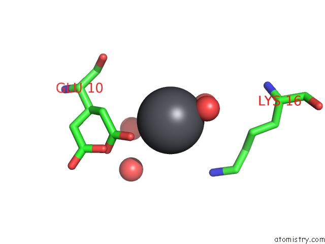
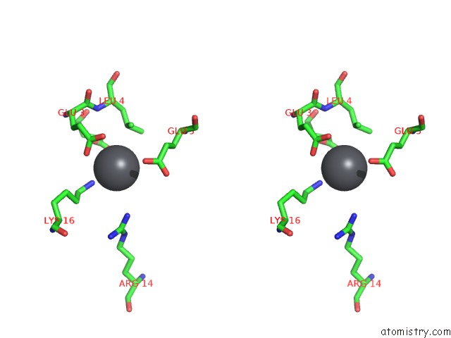
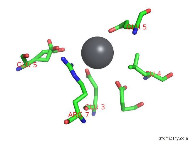
Lead binding site 1 out of 9 in 1hqj
Go back to
Lead binding site 1 out
of 9 in the Crystal Structure of A De Novo Designed Trimeric Coiled-Coil Peptide
Mono view

Stereo pair view
Mono view
Stereo pair view
A full contact list of Lead with other atoms in the Pb binding
site number 1 of Crystal Structure of A De Novo Designed Trimeric Coiled-Coil Peptide within 5.0Å range:
|
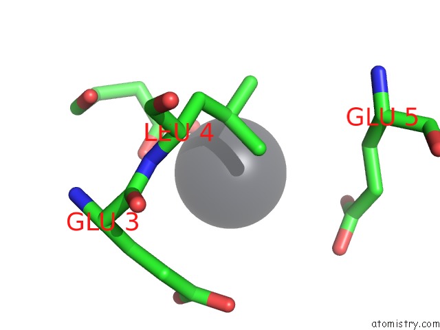
Lead binding site 2 out of 9 in 1hqj
Go back to
Lead binding site 2 out
of 9 in the Crystal Structure of A De Novo Designed Trimeric Coiled-Coil Peptide
Mono view

Stereo pair view
Mono view
Stereo pair view
A full contact list of Lead with other atoms in the Pb binding
site number 2 of Crystal Structure of A De Novo Designed Trimeric Coiled-Coil Peptide within 5.0Å range:
|
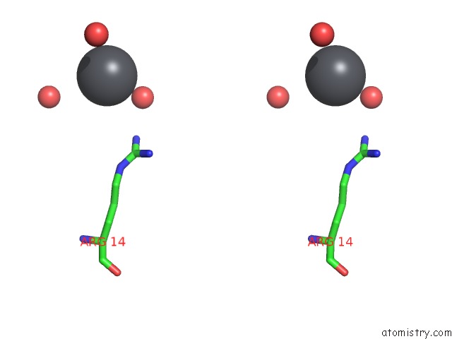
Lead binding site 3 out of 9 in 1hqj
Go back to
Lead binding site 3 out
of 9 in the Crystal Structure of A De Novo Designed Trimeric Coiled-Coil Peptide

Mono view

Stereo pair view
Mono view
Stereo pair view
A full contact list of Lead with other atoms in the Pb binding
site number 3 of Crystal Structure of A De Novo Designed Trimeric Coiled-Coil Peptide within 5.0Å range:
|
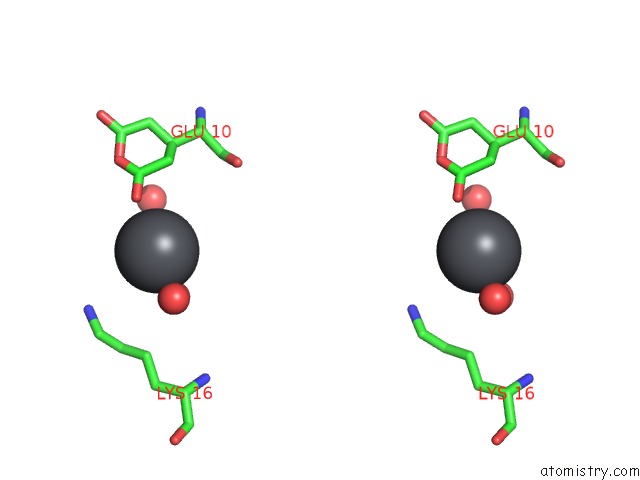
Lead binding site 4 out of 9 in 1hqj
Go back to
Lead binding site 4 out
of 9 in the Crystal Structure of A De Novo Designed Trimeric Coiled-Coil Peptide

Mono view

Stereo pair view
Mono view
Stereo pair view
A full contact list of Lead with other atoms in the Pb binding
site number 4 of Crystal Structure of A De Novo Designed Trimeric Coiled-Coil Peptide within 5.0Å range:
|
Lead binding site 5 out of 9 in 1hqj
Go back to
Lead binding site 5 out
of 9 in the Crystal Structure of A De Novo Designed Trimeric Coiled-Coil Peptide

Mono view
Stereo pair view
Mono view
Stereo pair view
A full contact list of Lead with other atoms in the Pb binding
site number 5 of Crystal Structure of A De Novo Designed Trimeric Coiled-Coil Peptide within 5.0Å range:
|
Lead binding site 6 out of 9 in 1hqj
Go back to
Lead binding site 6 out
of 9 in the Crystal Structure of A De Novo Designed Trimeric Coiled-Coil Peptide

Mono view

Stereo pair view
Mono view
Stereo pair view
A full contact list of Lead with other atoms in the Pb binding
site number 6 of Crystal Structure of A De Novo Designed Trimeric Coiled-Coil Peptide within 5.0Å range:
|
Lead binding site 7 out of 9 in 1hqj
Go back to
Lead binding site 7 out
of 9 in the Crystal Structure of A De Novo Designed Trimeric Coiled-Coil Peptide

Mono view
Stereo pair view
Mono view
Stereo pair view
A full contact list of Lead with other atoms in the Pb binding
site number 7 of Crystal Structure of A De Novo Designed Trimeric Coiled-Coil Peptide within 5.0Å range:
|
Lead binding site 8 out of 9 in 1hqj
Go back to
Lead binding site 8 out
of 9 in the Crystal Structure of A De Novo Designed Trimeric Coiled-Coil Peptide

Mono view
Stereo pair view
Mono view
Stereo pair view
A full contact list of Lead with other atoms in the Pb binding
site number 8 of Crystal Structure of A De Novo Designed Trimeric Coiled-Coil Peptide within 5.0Å range:
|
Lead binding site 9 out of 9 in 1hqj
Go back to
Lead binding site 9 out
of 9 in the Crystal Structure of A De Novo Designed Trimeric Coiled-Coil Peptide
Mono view

Stereo pair view
Mono view
Stereo pair view
A full contact list of Lead with other atoms in the Pb binding
site number 9 of Crystal Structure of A De Novo Designed Trimeric Coiled-Coil Peptide within 5.0Å range:
|
Reference:
P.Burkhard,
M.Meier,
A.Lustig.
Design of A Minimal Protein Oligomerization Domain By A Structural Approach. Protein Sci. V. 9 2294 2000.
ISSN: ISSN 0961-8368
PubMed: 11206050
Page generated: Thu Oct 10 09:59:35 2024
ISSN: ISSN 0961-8368
PubMed: 11206050
Last articles
Zn in 9J0NZn in 9J0O
Zn in 9J0P
Zn in 9FJX
Zn in 9EKB
Zn in 9C0F
Zn in 9CAH
Zn in 9CH0
Zn in 9CH3
Zn in 9CH1